Article Text

Abstract
Introduction Pancreatic exocrine insufficiency is a finding in many conditions, predominantly affecting those with chronic pancreatitis, pancreatic cancer and acute necrotising pancreatitis. Patients with pancreatic exocrine insufficiency can experience gastrointestinal symptoms, maldigestion, malnutrition and adverse effects on quality of life and even survival.
There is a need for readily accessible, pragmatic advice for healthcare professionals on the management of pancreatic exocrine insufficiency.
Methods and analysis A review of the literature was conducted by a multidisciplinary panel of experts in pancreatology, and recommendations for clinical practice were produced and the strength of the evidence graded. Consensus voting by 48 pancreatic specialists from across the UK took place at the 2019 Annual Meeting of the Pancreatic Society of Great Britain and Ireland annual scientific meeting.
Results Recommendations for clinical practice in the diagnosis, initial management, patient education and long term follow up were developed. All recommendations achieved over 85% consensus and are included within these comprehensive guidelines.
- pancreas
- pancreatic disorders
- pancreatic enzymes
- exocrine pancreatic function
Data availability statement
Data sharing not applicable as no datasets generated and/or analysed for this study. Not applicable.
This is an open access article distributed in accordance with the Creative Commons Attribution Non Commercial (CC BY-NC 4.0) license, which permits others to distribute, remix, adapt, build upon this work non-commercially, and license their derivative works on different terms, provided the original work is properly cited, appropriate credit is given, any changes made indicated, and the use is non-commercial. See: http://creativecommons.org/licenses/by-nc/4.0/.
Statistics from Altmetric.com
Introduction
Failure of the exocrine pancreas, referred to as pancreatic exocrine insufficiency (PEI), results in malnutrition, due to malabsorption of nutrients,1 2 the catabolic effects of the underlying pancreatic pathology and the impact of the disease on oral intake.3
Clinically, patients with PEI can present with non-specific gastrointestinal (GI) symptoms reflecting maldigestion, including steatorrhoea, weight loss, diarrhoea, abdominal pain and bloating.4 Consequently, patients may experience symptoms for prolonged periods before PEI is diagnosed.5
It is increasingly recognised that PEI adversely affects survival.6 7 Pancreatic enzyme replacement therapy (PERT) is the cornerstone of treatment and is associated with improved survival and quality of life (QoL) in patients with PEI.8–10
PEI may be underdiagnosed and undertreated in the UK, as demonstrated in other European countries.11 Patient support groups report management of PEI as the most common concern raised on their patient helpline (Pancreatic Cancer UK, 2015), and ‘difficulty in managing GI problems, diet and digestion’ are documented as the primary unmet need in patients with pancreatic cancer (PC).12 In addition, patients with chronic pancreatitis (CP) feel unsupported by healthcare professionals (HCPs) in the management of PEI (Pancreatitis Supporters Network, 2015). Consequently, there is a need for readily accessible, pragmatic advice for both specialist and non-specialist HCPs. The aim of this article is to provide evidence-based guidance on the diagnosis and management of PEI, including differential diagnosis and follow-up. This article does not make detailed recommendations regarding the management of cystic fibrosis (CF) as this is covered in depth in other guidelines and is outside the clinical expertise of the authors.
Methodology
PubMed literature searches were performed using the following core key words for each search: PERT OR pancreatin OR pancreatic exocrine insufficiency OR exocrine pancreatic insufficiency OR pancreas insufficiency OR pancreatic insufficiency. For each section topic, additional specific search terms were used (online supplemental table S1). English-language publications describing randomised controlled trials (RCTs), meta-analyses and observational studies were eligible for inclusion (cut-off date 22 May 2020; see online supplemental material). The review of the literature also identified other relevant articles, which were added if they met the inclusion criteria. The Grading of Recommendations Assessment, Development and Evaluation (GRADE) system13 was used to grade the strength of the recommendations (1=strong, 2=weak) and the quality of evidence (A=high, B=moderate, C=low). A consensus on the statements and GRADE scores was obtained from a voting panel of 48 pancreatic specialists, which included leading gastroenterologists, pancreatic surgeons, specialist dietitians, nurse specialists and research clinicians attending the 2019 Annual Meeting of the Pancreatic Society of Great Britain and Ireland annual scientific meeting). Statement 1.2 was added as a result of discussion within this meeting, and therefore was not subject to consensus voting.
Supplemental material
Results
Definition and diagnosis of PEI
Statement 1.1: PEI is defined as a reduction of pancreatic exocrine activity in the intestine at a level that prevents normal digestion (grade 1C; 100% agreement)
There is now agreement across pancreatic societies, globally, for this definition.14 15 The most common cause of PEI is CP due to loss of functioning pancreatic parenchyma and reduced secretion. Although PEI due to loss of functioning pancreatic parenchyma is also the result of pancreatic cancer, pancreatic resections and CF, other mechanisms exist. Decreased secretion can result from duct obstruction (eg, in periampullary tumour), or secondary causes, such as reduced endogenous stimulation (eg, in small bowel inflammation) and postprandial asynchrony (eg, after gastric surgery).16
Statement 1.2: Although the coefficient of fat absorption is regarded as the gold-standard diagnostic test for PEI, we recommend that the faecal pancreatic elastase (FEL-1) test is a suitable first-line test for PEI (grade 1B) (note this was not submitted for consensus voting)
Comments
A number of tests have been used for the diagnosis of PEI, including the coefficient of fat absorption (CFA), direct pancreatic function test (PFT), indirect 13C-labelled mixed triglyceride breath test and secretin injection at magnetic resonance cholangiopancreatography (sMRCP).17 18 These tests are not used in routine clinical practice in the UK,19 although some, particularly the CFA and direct PFT, are used in controlled clinical studies.18
FEL-1 is used in clinical practice as it is less invasive and readily available. FEL-1 is a measurement of a pancreatic exocrine-specific enzyme that is not degraded in the bowel lumen, is concentrated during intestinal passage and reflects the total overall pancreatic secretion. FEL-1 testing requires a small amount of faeces20 and is stable at room temperature for 3 days.21 A FEL-1 result of <200 µg/g stool suggests moderate PEI, while <100 µg/g suggests severe PEI. The sensitivity of an FEL-1 level of <200 µg/g for PEI, when tested in patients with known risk factors, has been shown to be 25%–65% in mild PEI; 33%–100% in moderate PEI and 82%–100% in severe PEI. The specificity of an FEL-1 level of <200 µg/g has been shown to be 55%–100% (although 6 out of 7 studies show specificity >90%).21–28
Statement 1.3: Stool samples for FEL-1 tests should undergo adjustment to standardised water content, when possible. A repeat test should be considered when a watery sample is reported by the laboratory, or when no definite cause of PEI is identified, to ensure correct diagnosis (grade 2B; 92% agreement)
False-positive results occur when water contaminates a specimen, such as in diarrhoea as samples can become diluted.29 If watery diarrhoea is reported, discussion with the laboratory is advised to ensure adjustment to a standard water content; one study showed that lyophilisation to a standardised water content of 75% was successful.29 If the cause of PEI is still not identified after further investigation and pancreatic imaging, stool testing should be repeated. As FEL-1 only tests for human elastase, the result is unaffected if the patient is taking PERT.21 The FEL-1 tests available in the UK were historically monoclonal,30 although more recently, polyclonal tests have become available and have been shown to have equal performance to the FEL-1 monoclonal tests.31
Statement 1.4: Positive markers of malnutrition, including clinical history, anthropometric measurements or serum micronutrient levels including magnesium, vitamin E and retinol-binding protein/vitamin A, can be used to support a diagnosis of PEI, if unclear. However, none of these markers should be considered in isolation when diagnosing PEI (grade 2A; 97% agreement)
A patient presenting with suspected malnutrition can be assessed in a number of ways. Initially, this should be conducted clinically by percentage weight loss; however, there is no agreed diagnostic percentage for PEI,3 32 33 and fluid balance is also important to consider. The Malnutrition Universal Screening Tool, or Nutritional Risk Screening 2002 if in hospital, are available. Although these tools are not specifically designed for PEI, they do have a high inter-rater reliability (κ=0.67–1.00) to identify patients that will likely benefit from nutritional support.34 35 A single body mass index (BMI) measurement is of limited diagnostic use on its own and can be normal in patients with PEI.36 37 BMI should also be interpreted with caution in an obese population.1 38 Evidence for recording specific anthropometric measurements is poor or absent for patients with PEI, with limited data in CP.1 39 Serial readings are of most use in this setting.
PEI is associated with some deficiencies in fat-soluble vitamins, trace elements and plasma proteins. Low levels of vitamin E have consistently been reported in patients with PEI or steatorrhoea caused by CP or CF.40–42 Studies have suggested retinol-binding protein (used as a marker of vitamin A status) may be lower in patients with alcohol-induced CP and steatorrhoea compared with patients with CP alone,41 although incidence studies in the CP population have not shown a significant difference between those with PEI and those without.43
Vitamin A deficiency night blindness is reported in case studies in patients with PEI.44 However, studies have not found that serum vitamin A deficiency is predictive of PEI.41 42 45 46
One study in patients with CP showed that serum magnesium <2.05 mg/dL was associated with PEI in univariate analysis; however, it only had a positive predictive value for PEI in CP of 0.58 (95% CI 0.39 to 0.75),43 while zinc deficiency has been associated with CP but not PEI specifically.47 Vitamin D deficiency has been shown to have a high incidence in CP (53%–66%) but no significant difference when PEI is present.1 41 45 48 Given the evidence available, no finding is specific enough to recommend using serum micronutrients alone as a diagnostic marker for PEI.
Statement 1.5: Cross-sectional imaging and an EUS (without secretin) cannot be used as a diagnostic test for PEI but can support its diagnosis and identify a cause. An abdominal CT scan should also be performed when PEI is diagnosed, especially to exclude a neoplastic cause (grade 2B; 94% agreement)
Comments
CT is recommended when investigating a clinical suspicion of CP, PC or other pancreatic disease that may cause PEI and similar symptoms.49–51 CT has been shown to differentiate CP from PC in 90.4% of cases and to exclude other aetiologies for pain or weight loss.52 Therefore, CT is the investigational tool of choice to exclude a pancreatic tumour as a cause when a diagnosis of PEI is made.53
Morphological changes of CP, including calcification and main pancreatic duct dilatation, can be identified on CT. Pancreatic calcification is a late or severe feature of CP,54 with PEI present in 50% of patients with substantial calcification.55 However, only 47% of patients with severe PEI and CP were shown to have significant morphological changes, such as atrophy and ductal dilatation on CT, in a study of 109 patients with CP.56 57 One study showed that duct dilatation, diagnosed via endoscopic retrograde cholangiopancreatography, has a stronger association with PEI (OR 5.8) than calcification (no effect).52 Parenchymal atrophy has been observed in 54% of patients with CP with CT58 and on magnetic resonance cholangiopancreatography (MRCP) in 20% of patients with PEI and 5% of patients without PEI in those with pancreatic pain symptoms, so is neither sensitive nor specific for PEI.59 In patients with CP undergoing MRCP without secretin, no significant difference in pancreatic duct size was observed between patients with or without PEI.60
Endoscopic ultrasound (EUS) can be used to identify very early parenchymal changes in non-calcific CP with a sensitivity of 84% and a specificity of 80%–100%, but a poor negative predictive value of 45% when compared with histology in high-prevalence groups, suggesting that histological changes can be overlooked.61–63 A single-centre experience has shown, in two separate studies in patients with CP (n=115 and 128; 30.4% and 37.5% with PEI, respectively), that both EUS markers and tissue stiffness measurements (strain ratio) show a linear increase in the likelihood of patients with CP having PEI.64 65 The probability of PEI, in the presence of calculi in the main pancreatic duct, was 80%, increasing to 82.8% if the main duct was also dilated. A further study comparing EUS findings with sMRCP showed reduced pancreatic function to be present in normal EUS exams and a predictive value of only 50% when six EUS criteria were present in a group with presumed pancreatic pain but an inconclusive or normal CT scan.66 Therefore, although radiological evidence of pancreatic morphological abnormalities is supportive of a diagnosis of PEI, further evidence, including maldigestion symptoms, evidence of malabsorption or a positive FEL-1 test, should be sought to confirm a diagnosis of PEI. A summary of key PEI diagnostic criteria is shown in box 1.
Diagnosis of PEI
PEI is highly likely with high benefit from PERT: no further test required as significant benefit from treatments and the negative predictive value of FEL-1 is not strong enough to prevent starting treatment
Head of pancreas cancer
Pre-surgery and post-surgery for head of pancreas cancer with or without pylorus preserving operation
Total pancreatectomy
Steatorrhoea or malabsorption symptoms in patients with CP with dilated pancreatic duct or severe pancreatic calcification
Severe necrotising pancreatitis
Patients that require initial investigation with FEL-1
GI symptoms of maldigestion in secondary care with or without known associated conditions
Maldigestion symptoms: steatorrhoea, weight loss, diarrhoea, abdominal pain or bloating
Associated conditions: patients with coeliac disease, IBS-D, HIV, type 1 diabetes and acute severe pancreatitis after initial phase
Following a positive FEL-1 test
A positive FEL-1 requires up-to-date cross-sectional imaging to exclude developing obstructive tumour or lesion as the cause
If subsequent investigation cannot find a morphological pancreatic cause of PEI, FEL-1 should be repeated even if patient has already started PERT
Perform a full malnutrition assessment with clinical history, and serum markers specifically including magnesium, vitamin E and retinol-binding protein
Where FEL-1 is normal in symptomatic patients with or without lower-risk associated condition and suspicion of maldigestion occurring.
When FEL-1 is low to provide a baseline for ongoing surveillance of micronutrient status
If micronutrient deficiency is demonstrated, other causes of malabsorption should be considered alongside PEI
In cases where there is significant weight loss or clinical signs of micronutrient deficiency to provide a basis for treatment
Aetiology of PEI
Statement 2.1: PEI is common in patients with CP (grade 2A; 95% agreement). Given the high prevalence of PEI in patients with severe CP, FEL-1 testing is not required and treatment is recommended in symptomatic patients (grade 2B; 95% agreement)
In CP, progressive destruction of the pancreatic tissue results in PEI.16 Steatorrhoea is a very late symptom that is associated with severe PEI and substantial decompensation, and is proposed to occur when >90%–95% of pancreatic parenchyma or function is lost.2 16 However, steatorrhoea was also demonstrated in patients with more than 10% secretory function,2 leading to concerns these publications might result in late diagnosis in patients with less severe disease.67 In the long-term, morphological changes, and the development of steatorrhoea or other malabsorption features, make PEI highly likely in patients with CP. Alcohol aetiology, pancreatic ductal obstruction, calcification and duration of disease have all been identified as contributing factors.68 PEI is reported in 94% of patients within 10 years of CP onset.37 Multivariate analysis has shown an association between increased mortality in CP and the presence of PEI (OR 2.59).6 Given the high probability of PEI in this setting, and the observed benefits of PERT, testing with FEL-1 is redundant and treatment with PERT is recommended.
Statement 2.2: PEI can occur following severe AP, especially in those with necrosis, recurrent AP or in the presence of pseudocysts (grade 2B; 100% agreement). Patients with acute necrotising pancreatitis should be routinely started on PERT once they are able to consume oral intake (good practice point (GPP); 92% agreement)
The FEL-1 test has been used to monitor the presence of PEI following AP. PEI prevalence, following severe and mild attacks of AP, has been reported as 60.5% and 55.6%, respectively.69 While there is some recovery in exocrine function over time, a meta-analysis of 10 initial and 39 long-term studies in patients with AP showed that PEI occurred at an overall rate of 62%, was higher in patients with severe AP (66%) compared with those with mild AP (46%) (p=0.001) and persisted in 35% of patients on long-term follow-up, with a pooled prevalence of 21% and 42% in patients with mild and severe AP.70 While PEI was more common in those with necrosis (p<0.0001), there was no difference in the prevalence of PEI in patients with ≥50% necrosis compared with that in patients with <50% necrosis (p=0.172).70 In a long-term follow-up study in patients who took part in an RCT comparing open necrosectomy with a step-up drainage technique at a median follow-up of 86 months (±11 months), the incidence of PEI was 29% in patients who had a step-up management of their necrosis compared with 56% in patients who underwent an open necrosectomy (p=0.03).71
Clinical judgement should be used when considering the likelihood of PEI in patients following severe AP, rather than a straightforward ‘indicated’ or ‘not indicated’ decision. A patient with almost total necrosis will most likely have PEI, whereas a patient who has recovered from solely oedematous pancreatitis, with no residual structural damage to the gland, may have normal exocrine function (although the prevalence of PEI appears to be 24%, even in this patient group).70 Testing after recovery can be helpful to determine whether long-term exocrine function is impaired. Where the clinician is unclear as to the likelihood or severity of PEI, PERT should be started to prevent nutritional compromise, and function tests should be requested.
Statement 2.3: PEI is common in both resectable and unresectable PC and is progressive in nature (grade 2A; 100% agreement). Given the high prevalence of PEI in this patient population, routine FEL-1 testing is not required, and treatment is recommended (grade 2B; 95% agreement)
PC induces a fibrotic reaction with a consequent reduction in exocrine function. Tumours occurring in the pancreatic head can directly obstruct the pancreatic ducts and are associated with PEI.72 PEI, gastroduodenal asynchrony and duodenal obstruction, are common in patients with unresectable PC, with PEI affecting 66%–94% of patients at presentation.73 74 Furthermore, PEI is progressive in patients with unresectable PC, with an estimated 10% reduction in exocrine function per month.74 The implication is clear: if a patient with PC is found to have normal pancreatic function at presentation, it is likely that PEI will subsequently develop. National guidelines advocate routine use of PERT in patients with unresectable PC,75 and pancreatic function tests are only needed where the cause of symptoms is unclear. However, despite this recommendation, three prospective randomised studies have reported a limited benefit of PERT in these patients.76–78
Surgery, particularly pancreaticoduodenectomy, is associated with PEI as it physically reduces the volume of the exocrine component of the pancreas and reduces the stimulation of the gland.79 80
In a meta-analysis of 693 patients with PC from nine observational cohort studies, the median prevalence of PEI was 44% before pancreaticoduodenectomy, 20% before distal pancreatectomy and 25%–50% in patients with locally advanced PC.81
The high prevalence of PEI in patients with PC (before and after surgery) means that little is gained from PEI testing. The benefits to patient survival (see Statement 3.1) and QoL (see Statement 3.2) with PERT, which have been demonstrated in these patients, means that treatment would usually be indicated, regardless of the FEL-1 result.75
Less common causes of PEI
Statement 2.4: Patients with diabetes mellitus may have PEI; however, the exact prevalence is not clear. Those with relevant symptoms should be offered PERT and investigated for a pancreatic pathology (grade 2C; 94% agreement)
There are few large studies investigating the prevalence of PEI in patients with diabetes mellitus (DM), but estimates of 26%–57% and 12%–36% have been reported for patients with type 1 and type 2 DM, respectively.82 Low FEL-1 levels have been associated with reduced glycaemic control in patients with DM (p=0.031),83 and one study also reported a weak association with reduced BMI.84 Another study reported a negative correlation between FEL-1 level and duration of DM.83 These findings further support the concept that PEI may be a complication of DM.
One study in patients with DM and low FEL-1 (defined as <100 µg/g) showed that PERT reduced the frequency of episodes of hypoglycaemia.85 This study, however, included few patients with symptoms that would have been compatible with PEI.
Despite a high prevalence of PEI in patients with DM, the identification of individuals who would benefit from PERT, and the potential long-term effects on important diabetes-related outcomes, requires further investigation as PEI should be considered a symptom of a disease rather than a disease in its own right.
New onset diabetes in the absence of typical risk factors should be investigated as this may be the first sign of pancreatic cancer or CP.86
Statement 2.5: PEI should be considered in patients with symptoms of maldigestion in the absence of severe pancreatic disease. In these cases, FEL-1 testing should be undertaken (grade 2B; 100% agreement)
FEL-1 has been successfully used as a case-finding tool for PEI in a minority of patients with coeliac disease, Type 1 DM, HIV, diarrhoea-predominant irritable bowel syndrome (IBS-D) and difficult symptoms after bariatric surgery (table 1). FEL-1 levels<200 µg/g stool have been reported in 11.5%–13.1% of patients presenting to secondary care clinics, and a separate study demonstrated that 79.6% of patients with low FEL-1 responded to PERT,87 88 indicating that they have PEI. Follow-up data are required to ascertain how many of these patients developed pancreatic pathology.
Prevalence of PEI, potential value of PERT and recommendations for less common causes of PEI
Recommendations for other, less common, causes of PEI, such as coeliac disease, HIV, IBS-D and alcohol-related liver disease, are shown in table 1, and detailed supporting evidence is shown in the online supplemental material.
The value of PERT
Statement 3.1: PERT is associated with improved survival in patients with CP (grade 1C) and PC (grade 2B). Additionally, PERT is associated with improved nutritional status in patients with CP (grade 1C) and PC (grade 1C) (95% agreement)
PERT has been shown to improve symptoms and digestion in patients with pancreatitis.89 In patients with AP, PERT maintained global health status.90 Among patients with CP, PERT has been shown to improve fat and nitrogen absorption, nutritional parameters and GI symptoms.91
PEI has been shown to be an independent factor related to mortality in patients with CP,6 and an absence of PERT on discharge was an independent risk factor for survival in those undergoing surgery for CP.92
In a small, randomised trial conducted over an 8-week period in 21 patients with unresectable PC, those receiving PERT maintained body weight (gain 1.2%; 0.7 kg), while patients without PERT lost body weight (loss 3.7%; 2.2 kg) (both p=0.02). CFA increased by 12% in the PERT group and declined by 8% in the control group, but this difference did not reach statistical significance (p=0.13).77 An RCT in Korea failed to observe any benefit of PERT on body weight (p=0.381), subjective global assessment (p=0.18) or survival (p=0.744) compared with placebo over an 8-week period in 67 patients with unresectable PC.76 It should be noted that patients were not permitted to receive H2-receptor antagonists or proton-pump inhibitors (PPIs) and, consequently, PERT may not have been effective without adequate neutralisation of gastric acid. The dose of PERT was also quite low at 2×25 000 units of lipase three times daily with main meals and one capsule with snacks. In addition, only 43% of patients included had severe PEI (FEL-1 level <100 µg/g stool), and only a third having tumours in the pancreatic head (35.5% in the PERT group; 33.3% in the placebo group). Furthermore, although the difference was not statistically significant, more patients in the PERT group had a FEL-1 level ≥200 µg/g than those in the placebo group (44.1% vs 21.2%; p=0.13). Hence, this trial has several limitations, and the results should be interpreted accordingly.
Further work in exploring the potential benefits of PERT on survival identified a link between the severity of PEI and reduced survival in a non-randomised study of 194 patients with PC. A FEL-1 level ≤20 µg/g was an independent predictor of survival (HR 1.59; p=0.023).7 Treatment with PERT appeared to improve survival in a recent Spanish retrospective single-centre study.9 Patients (n=160) with unresectable PC were treated by different teams who either prescribed high-dose PERT, if needed, or did not. Median survival was significantly longer among patients managed by the team more likely to prescribe PERT (189 days; 95% CI 167.0 to 211.0 days vs 95.0 days; 95% CI 75.4 to 114.6 days) (HR 2.12; 95% CI 1.49 to 3.00; p<0.001). However, the results should be interpreted with caution due to potential selection bias arising from the retrospective non-randomised study design. In a retrospective study of 469 patients who had undergone resection of periampullary cancer, PERT was associated with a median increase in survival on both multivariate (p=0.044) and propensity-matched analyses (p=0.009). Survival benefit was most markedly observed among patients treated with PERT and with pancreatic duct widths of ≥3 mm (HR 0.64; 95% CI 0.47 to 0.89; p=0.006).8
A retrospective, population-based, propensity-matched, observational study in the UK matched 1614 patients with PC who did or did not receive PERT.8 The adjusted median survival time was 262% longer among PERT-treated patients compared with matched, non-PERT-treated controls (survival time ratio 2.62; 95% CI 2.27 to 3.02). This survival benefit was present in patients who did not receive chemotherapy or surgery, suggesting that this benefit was independent of other interventions.
Overall, there are conflicting data regarding PERT and survival, with RCTs limited by heterogeneous populations and other data limited by retrospective or observational trial design. More robust studies are required to strengthen this recommendation.
Importantly, a large survey of 208 patients with PC who were prescribed PERT in the USA identified that only 89 patients (66% of those prescribed PERT correctly), were taking their enzymes as instructed. Predominantly, this was due to incorrect prescriptions, with 35% of patients not prescribed PERT with meals and snacks. Patients who were taking their enzymes with meals, rather than before or afterwards, were more likely to gain weight (p=0.022) and demonstrate less maldigestion (p=0.003) and less steatorrhoea (p=0.04) compared with those not taking PERT correctly.93 The high incidence of incorrect timing is an important consideration when analysing data from studies assessing the clinical response to PERT, especially when data are collected retrospectively.
Statement 3.2: Treatment with PERT improves QoL in patients with PEI (grade 1C; 100% agreement)
While PERT is useful for treating PEI, studies that address whether it has a positive effect on the QoL of those taking it have been of variable quality. Achieving equipoise, and assessment over an appropriate time period, can affect study design and interpretation, especially in the PC cohort.
Being able to justify the pill burden associated with PERT by confirming an associated improvement in QoL is both relevant and important. While initiating PERT often requires specialist input, there is an ongoing need for patient self-management of PERT dosing. A small qualitative trial explored patient experiences, recruiting patients who were receiving PERT for at least 6 months following pancreaticoduodenectomy for malignancy. Patients reported concerns over the use of PERT at social events, difficulties in remembering to take treatment and struggles with dose adjustment. Socially debilitating symptoms of PEI (excessive flatulence and steatorrhoea), and the importance of PERT, were also acknowledged. The authors concluded that education and access to specialist support was important to patients.94
Two cohorts of patients with PEI, secondary to CP, were observed over a 1-year period, with assessment at 6 months and 1 year. Assessment of symptoms and QoL in patients already receiving PERT (n=206) and in patients starting treatment with PERT (n=88) were compared. PERT dosing was not controlled by the study design. Data from 294 patients were analysed; 256 patients completed the validated Gastrointestinal Quality of Life index (GIQLI). GIQLI scores in the untreated were significantly lower than those on treatment at baseline (p<0.001), but this difference disappeared after 6 months of treatment. The mean total GIQLI score improved significantly over 1 year for the study population (p<0.001), as did all five subscores (symptoms scale, physical function, social function, emotion and medical treatment). Overall, both cohorts exhibited significant improvements in QoL (both p<0.001), suggesting that improvement in QoL may be sustained with continued PERT.10
Another prospective study examined the effect of PERT versus placebo on QoL in patients with moderate-to-severe AP, using the Clinical Global Impression of Disease Symptoms (CGIDS) tool alongside version 4 of the validated Functional Assessment of Cancer Therapy-Pancreas QoL questionnaire.90 Recruitment issues led to termination of the study with 56 patients enrolled, but over the 26–30 day study period, there was an improvement in the CGIDS scores with numerically, but not statistically significant worse flatulence in the placebo group (p=0.503). Of note, was a FEL-1 level <200 µg/g in only 20 patients. The QoL assessment also favoured the PERT group, but only the ‘emotional well-being’ score, demonstrated a significant improvement in the PERT group versus placebo (p=0.04).90
More recently, work has been undertaken to develop a tool to identify patient-reported outcome measures in patients with CP (n=91). This tool correlated well with GIQLI scores, and patient scores correlated with disease severity (p<0.0001). It could be used in future research to provide a less invasive measure of treatment effect.95
In summary, there are data supporting the use of PERT to improve QoL in patients with AP, CP and resectable and non-resectable PC, but further high-quality studies are required.
Management of PEI
Statement 4.1: Patients should consent for the porcine nature of PERT (GPP; 97% agreement)
All currently available PERT preparations are porcine (a non-porcine PERT formulation was in development, but it failed to meet its primary endpoint in a phase III clinical trial).96 Patients should be made aware of the porcine origin of PERT before commencing therapy. Jewish and Muslim faith leaders consent to the use of PERT as there are no alternatives available.97
Statement 4.2: PERT should be started at a dose of at least 50 000 units lipase with meals and 25 000 units lipase with snacks (grade 1A), and patients should be encouraged to adjust their dose if this is ineffective (grade 2C) (92% agreement)
The choice of PERT preparation needs to consider the lipase units the patient requires and the number and size of capsules the patient is able to swallow. Studies support the use of at least 50 000 units lipase as a suitable starting dose with meals and 25 000 units lipase with snacks.91 98 All guidelines endorse dose escalation if the initial dose is not effective.77 99 100
Statement 4.3: Consumption of pancreatic enzymes should be spread out throughout a meal (grade 1C; 100% agreement)
A study examining the timing of PERT in 24 patients with CP suggested that spreading the dose throughout the meal supported optimal absorption.101 However, this trial was limited by strict exclusion criteria: no pancreatic or GI surgery, no antiacid therapy, no gastro paresis, no excessive alcohol intake or malignancy, no opiate prescription and the use of a prokinetic prior to the test.101 These data were supported by a survey of 262 patients, which demonstrated that patients consuming PERT throughout meals experienced fewer abdominal symptoms and more weight gain compared with patients who consumed PERT before or after meals.93
Statement 4.4: If capsules cannot be swallowed, they should be opened, placed on an acidic puree and swallowed at intervals throughout the meal, and the mouth should be rinsed with cool water to prevent ulceration (GPP; 97% agreement)
Some patients are unable to swallow capsules. In those circumstances, the capsules can be opened and the contents taken on a spoonful of cold acidic food, such as fruit puree, apple sauce, jam or fruit yoghurt and swallowed immediately.
This should then be washed down with a cool or room-temperature drink to ensure that no granules remain in the mouth, where they could cause ulceration. Special care should be taken by those who wear dentures. Granules should not be chewed or crushed as this removes the enteric coating, resulting in premature activation of the enzymes.
Statement 4.5: There is no maximum dose of PERT in adults (GPP); however, where doses exceed 100,000 units lipase with meals, comorbidities should be excluded (GPP; 91% agreement)
The most frequently documented side effects of PERT are nausea, vomiting and abdominal discomfort. Further details of rarer side effects can be found in Statement 7.1.
The product itself does not raise toxicity concerns in the event of unnecessary or excess treatment. However, pill burden should be considered, and when dose escalation has not resolved ongoing symptoms, comorbidities should be considered (see Statement 5.1).
Statement 4.6: Dietary fat restriction is not routinely recommended, but very high-fibre diets should be avoided (grade 1C; 96% agreement)
Some patients require a high-fat, high-protein diet if they are nutritionally depleted and have a low appetite. Increasing the fat content of the diet has been shown to be well tolerated in patients with CP.102 103 Low-fat diets may exacerbate malnutrition and should be avoided, unless there is a specific reason for fat intolerance. Very high-fibre diets (>25 g/day) may absorb enzymes and delay nutrient absorption and, therefore, are not recommended.104
Statement 4.7: Where needed, enteral feeds should be peptide and medium-chain triglyceride-based (grade 2C; 100% agreement)
One small pilot study examined the use of peptide versus polymeric feeds in patients with severe AP.105 Nutritional status and biochemical markers were assessed 2 weeks into the study. In populations with severe AP, it is unlikely that any significant change would be observed within this time period. While the study concluded that there was no difference between the two feed types, analysis of the secondary endpoints, including weight loss and length of stay, demonstrated an improvement in the peptide-fed group, suggesting that malabsorption of feed, secondary to PEI, has an impact on recovery.105 Other guidelines have used extrapolated data from studies comparing enteral and parenteral nutrition to make strong recommendations regarding the use of peptide-based feeds (grade A), but these data support enteral over parenteral nutrition, rather than a specific feed,106 resulting in conflicting recommendations.107
Statement 4.8: Both acid (grade 1C) and temperature (GPP) impact the efficacy of PERT. The addition of a PPI can be useful if PERT is not effective (grade 1C, 97% agreement). If one PERT product remains ineffective, despite the use of a PPI, a trial of an alternative PERT preparation is recommended (GPP)
Several studies have shown increased efficacy of PERT with the addition of a PPI, as PERT may be inhibited by gastric acid.108–110 However, the data are not consistent, with some studies showing no benefit.111 112 Specifically in pancreatic cancer, one study suggests a PPI may help reduce further pancreatic atrophy,113 but studies are limited by heterogenous cohorts.110 111
Given the conflicting data, and the potential for long-term complications of PPI use, including Clostridium difficile infection, hypomagnesaemia, nausea and diarrhoea,114 115 the addition of a PPI is a second-line treatment for the management of PEI.
Optimum storage of enzymes (below 25°C) to prevent deterioration of function during storage is important.116–119 Each PERT product functions slightly differently at different pHs and timings,120–122 so considering the use of an alternative PERT product may be beneficial, particularly in cases where gastric transit is altered.
Statement 4.9: Patients with PEI receiving enteral feeds usually tolerate semielemental (peptide) preparations. However, where malabsorption symptoms persist, enzymes can be flushed via the feeding tube every 2 hours or added to the feed itself (GPP; 93% agreement)
Semi-elemental (peptide) feeds require less PERT to achieve complete lipolysis than polymeric feeds123; therefore, peptide feeding is recommended for patients with PEI who are receiving enteral feeds. When considering a choice of feed product, it is sensible to consider the total fat content and the proportion of medium-chain and long-chain triglycerides as well as the polysaccharide content. This information, alongside nutritional requirements, fluid needs and tolerance, will assist in the selection of an appropriate feed. Effective mixing of feed with enzymes is difficult to achieve, especially with jejunal feeding, and administration of the enzymes, either as flushes or mixed directly with the feed, has been documented,124 125 but evidence is limited, with no safety or stability data available (box 2). Jejunal feeding will, theoretically, reduce stimulation of the pancreas126 and may mean that patients are more likely to require PERT compared with those receiving gastric feeding.
Administration of PERT with enteral feeds
Powdered enzymes and feeding tubes
NB. Once mixed, use all products immediately. Do not leave to stand
Giving PERT as flushes: mix 1 g scoop pancreatin powder (Pancrex V Powder, Essential Pharmaceuticals, UK) with 50 mL sterile water. Shake well and immediately flush via a feeding tube. Do not give with other medication. Do not flush between the feed and the enzyme as this will reduce the mixing of the feed with the PERT. Administer every 2 hours hours throughout enteral feeding, increase dose of PERT if needed.
Mixing PERT with feed: add 1–2 g Pancrex V Powder directly to the feed in a feeding reservoir. Shake well. Hang straight away and for 4 hours hours only, increase dose of PERT if needed. (NB. Some feeds congeal when PERT is added - discuss with a tertiary centre dietitian prior to adding PERT to feed)
Flushing granules/mini-microspheres via large bore tubes (>CH20): mix with an acidic juice (such as Fortijuce (Nutricia Clinical Care, UK); Ensure Plus Juce (Abbott Nutrition, UK); Fresubin Jucy Drink (Fresenius Kabi, UK)) and flush via the feeding tube every 2 hours hours throughout enteral feeding, increase dose of PERT if needed
PERT, pancreatic enzyme replacement therapy
Statement 4.10: When malabsorption has been corrected, nutritional status and bowel symptoms should improve; thus, monitoring of these symptoms should support ongoing treatment or dose escalation (GPP; 95% agreement)
To assess the need for PERT dose escalation, nutritional and symptom assessments can be used as summarised in table 2.
Evaluation of the need for PERT dose escalation: nutritional and symptom assessments
Differential diagnosis/treatment failures
Statement 5.1: When symptoms continue, despite treatment with pancreatic enzymes, clinicians should ensure that PERT products are being taken correctly, the dose has been adequately escalated and other conditions have been excluded (GPP; 100% agreement)
Treatment with PERT does not completely normalise digestion, and undertreatment is common, with many patients continuing to experience symptoms of malabsorption, placing them at risk of nutritional deficiencies. A study in Germany showed significant improvement in GI symptoms, pain and QoL in both newly diagnosed patients and those with established CP, following review of PERT (p<0.001), demonstrating the benefit of dose review.10
No treatment is 100% effective, but the steps in box 3 should help to optimise management of PEI. If symptoms of PEI continue, despite optimising PERT dose, then other causes should be considered. Small bowel bacterial overgrowth (SBBO) was reported in 36% of patients with CP in a meta-analysis of nine studies.127
Practical guidance for the management of continued symptoms despite commencement of PERT
Timing and dosing
Ensure that the correct dose of enzymes is prescribed; minimum of 50 000 units lipase per meal and 25 000 units lipase per snack. Many patients require more than this for adequate digestion. Escalate the dose and monitor for effect
Check PERT is taken appropriately, that is, with all food and milky drinks, spread throughout the meal rather than taking at the start or end of the meal
Ensure PERT is being taken with all intake, for example, nutritional supplements, milky drinks, eating outside the home, snacks
Preventing denaturing of the enzyme
Check how the PERT is stored (<25°C: avoid storage in direct sunlight, cars, near heat sources such as kettles)
Consider gastric acid suppression
Factors affecting interpretation of symptoms
Exclude constipation and overflow diarrhoea
Check if any other medication that may be causing abdominal symptoms is being taken, for example, laxatives, chemotherapy, metformin, antiemetics
If symptoms are not controlled
If symptoms remain despite a dose of ≥10 000 units lipase/kg/day, then other causes of the symptoms should be considered before increasing the dose of PERT further
Other causes of loose stools should be investigated, for example, SBBO, IBD, BAM, infection, coeliac disease, lactase deficiency, other food intolerances (see online supplemental table S2 for further details)
Bile acid diarrhoea, sometimes referred to as bile acid malabsorption (BAM) should also be considered; however, while the prevalence of this condition in patients with PEI is unknown, many patients with pancreatic disease have undergone cholecystectomy as part of the management of gallstone disease,128 which is associated with a risk of BAM.129 130
Two studies have shown an increased prevalence of PEI in patients with IBS-D,131 132 but the prevalence of functional symptoms in patients with exocrine dysfunction is unknown.
In summary, once dose is optimised, ongoing symptoms should be investigated by taking a structured history and performing coeliac serology, investigation for SBBO using a glucose hydrogen breath test, colonoscopy with colonic biopsies and a selenium homocholic acid taurine (SeHCAT) scan. Other causes of ongoing symptoms should be investigated as per the British Society of Gastroenterology guidelines for the investigation of chronic diarrhoea in adults.133
Long-term follow-up in patients with PEI
Statement 6.1: atients with PEI often have ongoing problems with malnutrition, malabsorption and bowel symptoms; therefore, long-term follow-up is recommended (GPP; 100% agreement)
Long-term support is not easily accessible for patients with pancreatic disease in the UK, with a survey of UK practice demonstrating that most patients were not followed up after discharge,134 resulting in inconsistent advice, often provided by non-specialists. A lack of guidance on the management of GI symptoms, diet and digestion has been identified as the primary unmet need of both patients and their carers.12 In benign disease, GI symptoms may have a long-term impact on QoL in those with AP and CP.135 136
Long-term follow-up should address issues with education, the management of PERT and should provide formal provision for long-term monitoring of nutritional markers and micronutrient status.137 This nutritional follow-up should include clinical, anthropometric and biochemical measures of nutritional status,137 138 and the development of a structured annual assessment (figure 1) could follow the model established for coeliac disease.138 Clinicians need to be pragmatic; the assessment of patients with rapidly progressing disease should focus on QoL and management of symptoms, whereas long-term health must be a consideration in those with benign disease or those who have undergone treatment with curative intent (table 3).

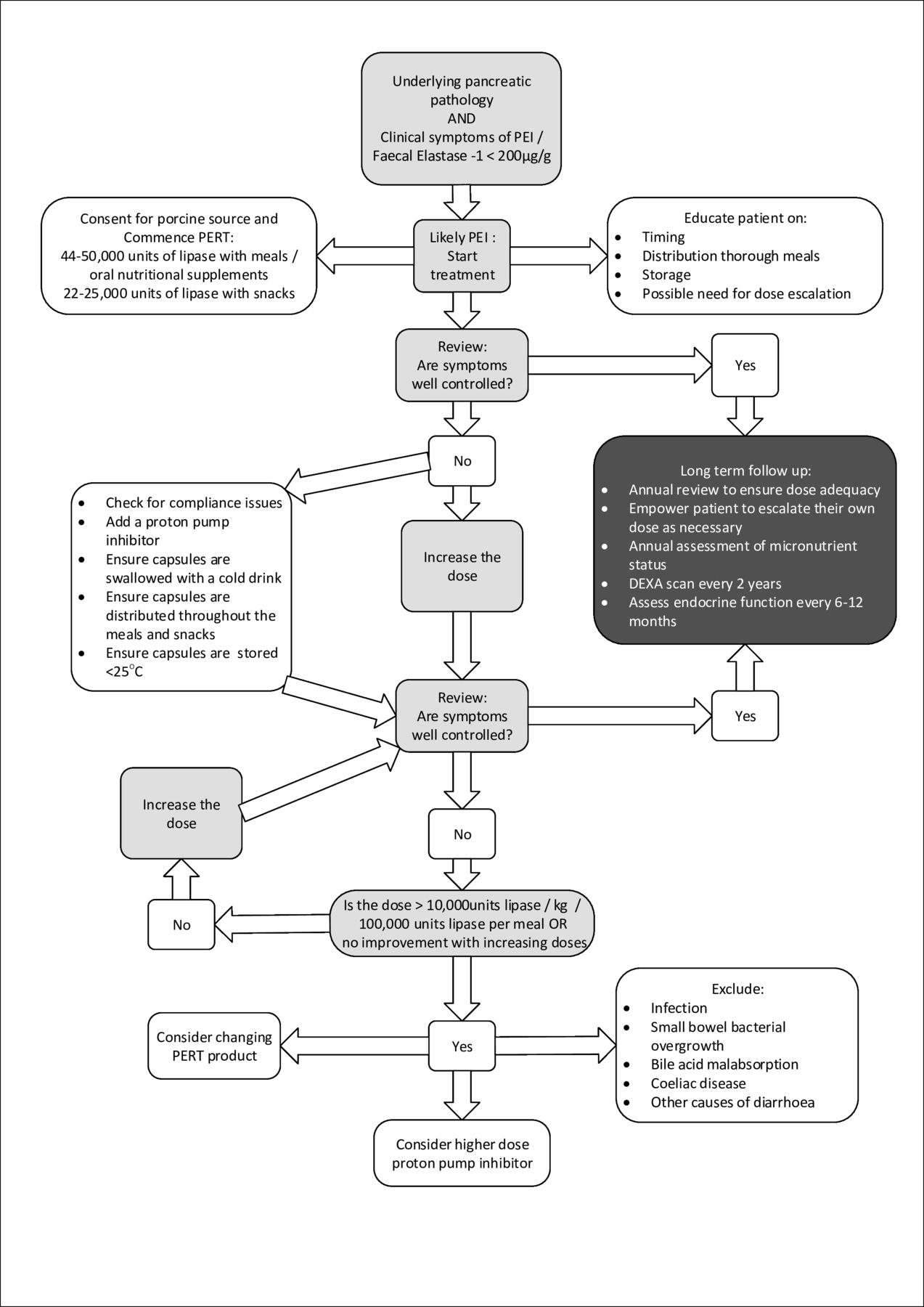
Summary of guidance. DXA, Dual-energy X-ray absorptiometry; PEI, pancreatic enzyme insufficiency; PERT, pancreatic enzyme replacement therapy; PPI, proton-pump inhibitors.
Long-term follow-up recommendations
Statement 6.2: Based on the literature search, no studies have been published to support a direct association between PEI and bone health. However, there is a high prevalence of osteoporosis and osteopenia in patients with pancreatic diseases that also result in PEI. Therefore, the best practice would be to implement routine monitoring of bone mineral density in pancreatic diseases (GPP; 100% agreement)
Limited data are available to support a conclusive link between PEI and bone health, and no studies were identified from the literature search that investigated a direct association (see details in the online supplemental material).
Min et al139 reported that 31/45 (68.9%) patients with CP and PEI had either osteoporosis or osteopenia. A statistically significant association between low FEL-1 and low bone mineral density was reported in patients with PEI and CP, as assessed by X-ray (p<0.05), but not by dual X-ray absorptiometry (DXA)—the latter being the gold standard for assessing bone mineral density.140 Patients on PERT had higher bone mineral density, as assessed by DXA (p<0.05). In 167 patients with osteoporotic bone fractures, it was found that PEI (low FEL-1) was more frequent compared with non-fracture controls141: 34% of patients with osteoporosis had FEL-1 levels<200 µg/g, and FEL-1 levels were 65% lower in patients than in healthy controls. Furthermore, vitamin D, parathyroid hormone and calcium levels correlated with FEL-1 level (p<0.01).139
Finally, in an uncontrolled study, 13 of 14 patients with CP, all of whom had PEI, had either osteoporosis or osteopenia.142 No association was identified between osteoporosis/osteopenia and the secretin test or faecal fat excretion.
A systematic review and meta-analysis of the prevalence of osteoporosis and osteopenia in CP identified eleven studies and 10 were included in the meta-analysis: seven articles and three conference proceedings. This meta-analysis of 513 patients found that 23.4% of patients with CP had osteoporosis, 39.8% had osteopenia and 65% had either osteoporosis or osteopenia.143 Only one of the studies included in the systematic review by Duggan et al. (2014a) was retrieved in the broad literature search for the current systematic review,142 and none were retrieved following the application of search filters. Of the 11 studies in the Duggan et al. (2014a) review, five144–148 reported an association between low bone mineral density and low FEL-1, while one study149 found no such association.
The National Institute for Health and Care Excellence guideline for pancreatitis137 recommends bone mineral density assessment, by DXA scan, every 2 years for those with CP.137 The HaPanEU guidelines150 recommend that, as well as regular DXA scanning, all patients with CP should receive basic preventative measures to protect against the deterioration of bone mineral density, including adequate diet (particularly calcium and vitamin D intake), regular weight-bearing exercise and the avoidance of smoking and alcohol. For pancreatic diseases other than CP, given the morbidity and mortality associated with osteoporosis and fracture, as well as the preventable nature of osteoporosis, it seems prudent to recommend routine monitoring of bone mineral density using DXA scans in benign or curative disease.
Adverse events
Statement 7.1: PERT is not associated with any significant complications (grade 1A; 100% agreement)
There are many RCTs examining the efficacy of PERT, and all report safety data for the duration of the trial. Some of these are open-label extensions, up to 12 months, but there is a paucity of RCTs examining the long-term effects of PERT.
Industry-funded studies report mild abdominal pain as an adverse event (AE) in patients with CP (3/34) but no patients withdrew as a result of an AE.151 Whitcomb et al. identified 8/25 patients reporting AEs (flatulence, change in bowel habit, increased bowel frequency, hypoglycaemia and hyperglycaemia) in a trial exploring the use of PERT in a mixed cohort of patients with CP, or pancreatic resection, who were taking 72 000 units lipase with meals and 36 000 units lipase with snacks.152 Interestingly, there were 8/29 AEs of similar nature in the placebo group.
A 6-month open-label extension in 48 patients already established on PERT due to CP or pancreatic surgery found four patients reported AEs potentially associated with treatment: weight gain (n=1), abdominal distension (n=1), flatulence (n=1), abdominal pain (n=1) and diarrhoea (n=1). These were all classified as mild, and there were no withdrawals from the study as a result.153
In a cross-over trial using two different PERT products, the total number of AEs across both groups was reported as: headaches (4/46), diarrhoea (2/46), infection (3/46) and abdominal pain (2/46).99 Subsequently with an additional cohort 8/61 patients reporting AEs including constipation, abdominal discomfort and frequent bowel motion.91
A placebo-controlled study examining the use of PERT in 95 patients with type 1 DM found no difference in the frequency of AEs between the intervention group (40 000 units lipase with meals and 20 000–30 000 units lipase with snacks) and the control group; however, a reduction in mild and moderate hypoglycaemia was observed in the intervention group at the end of the study.85 The most common AEs reported were headache, infection, pain, diarrhoea and dyspepsia; the incidence of each AE was not specified, but the overall incidence was the same in both the intervention and placebo groups. Changes in glycaemic control, due to PERT in patients with existing PEI, were only reported as sufficient to cause a change in management in one patient in a cohort of 36 patients with DM as part of a larger study consisting of 54 patients.154 There was no treatment-related AEs in the non-diabetic group.154
While these trials are small, they are all consistent in results. Regardless of trials not reporting serious AEs, fibrosing colonopathy (FC) cannot be ignored.
Statement 7.2: There is limited evidence to suggest that long-term use of very high-dose PERT may be associated with fibrosing colonopathy in children with CF, resulting in a dose restriction of 10 000 units lipase/kg/day in this population (grade 1C; 100% agreement)
Comments
Cases of FC were first reported in 1992 and are thought to be associated with high doses of PERT (>19 000 units lipase/kg/day in the initial reports).155 Causal factors, including the presence of Eudragit L30D-55 acrylic resin within the enteric coating, were debated,156 but the initial research into this was discredited.157 Contributing factors were summarised as young age, previous intestinal surgery, meconium ileus and the use of corticosteroids, deoxyribonuclease and H2 blockers, and all cases were reported in children with CF.158–160
Patients who presented before the formation of strictures were identified as having reversible symptoms that resolved on lowering the dose of PERT, suggesting the risk increased with higher doses. However, following the publication of case reports describing the presentation of FC in neonates who had not received any PERT, the debate over the mechanism behind the development of FC continues.160 161 At present, a maximum dose of 10 000 units lipase/kg/day is recommended for children with CF. There is no maximum dose proposed for adults or children with non-CF-related PEI. Recognition features of FC are documented in table 4.
Symptoms of fibrosing colonopathy
Use in pregnancy and breastfeeding
Statement 8.1: Further work is required to confirm the efficacy and safety of PERT during pregnancy and breastfeeding; although the authors have experience of successful use in pregnancy, the numbers are small and there are no published data. Malabsorption should be avoided in pregnancy (GPP; 93% agreement)
The systematic literature search did not identify any articles concerning the use of PERT in pregnancy or breast feeding. Essential fatty acids are required for brain and retinol development in the first 8 weeks of gestation.159 Thus, it is important that adequate PERT is maintained in pregnancy.
Summary
This document serves as an evidence-based guide for the diagnosis and treatment of PEI. Data were derived through a pragmatic review of the literature, and conclusions were assessed by a multidisciplinary panel of experts in pancreatology at the 2019 Annual Meeting of the Pancreatic Society of Great Britain and Ireland.
The strengths of this work are the comprehensive nature of the assessment and the focus on the practical aspects of manging PEI. Also addressed are areas of uncertainty, or areas where practice is difficult or variable, such as diagnostic tests and treatment of patients whose symptoms are vague or refractory to PERT.
Data availability statement
Data sharing not applicable as no datasets generated and/or analysed for this study. Not applicable.
Ethics statements
Acknowledgments
The authors would like to acknowledge the support of Jeremy French and Lynne McCullum in developing the content of this document.
References
Supplementary materials
Supplementary Data
This web only file has been produced by the BMJ Publishing Group from an electronic file supplied by the author(s) and has not been edited for content.
Footnotes
Contributors All authors were involved in the design, writing and reviewing of this document. All authors contributed to the design, analysis and interpretation of data. All authors drafted sections of the paper and critically appraised the entire paper prior to submission.
Funding Editorial assistance was provided by Alpharmaxim Healthcare Communications which was funded by Mylan Products Limited. The guideline content is independent of and not influenced by Mylan. Publication fees were met from an academic research account affiliated with Sheffield Teaching Hospitals.
Competing interests MEP has received honoraria for teaching and resource development from Mylan, Nutricia, and Sanofi. AH has received honoraria for teaching and resource development from Mylan. JSL has received honoraria for teaching and consulting from Mylan. KJR has received honoraria for teaching from Mylan and Abbott. LM has received honoraria for teaching and conference attendance from Mylan. SND has received honoraria for teaching, consulting and conference attendance from Mylan and Abbott. RK has received honoraria for teaching from Mylan.
Provenance and peer review Not commissioned; externally peer reviewed.
Supplemental material This content has been supplied by the author(s). It has not been vetted by BMJ Publishing Group Limited (BMJ) and may not have been peer-reviewed. Any opinions or recommendations discussed are solely those of the author(s) and are not endorsed by BMJ. BMJ disclaims all liability and responsibility arising from any reliance placed on the content. Where the content includes any translated material, BMJ does not warrant the accuracy and reliability of the translations (including but not limited to local regulations, clinical guidelines, terminology, drug names and drug dosages), and is not responsible for any error and/or omissions arising from translation and adaptation or otherwise.