Article Text

Abstract
Objective Functional dyspepsia (FD) is an extremely common functional gastrointestinal disorder, the pathophysiology of which is poorly understood. We hypothesised that impaired intestinal barrier function is involved in the onset and persistence of this disorder by inducing low-grade inflammation. Therefore, our aim was to evaluate duodenal mucosal integrity and low-grade inflammation in patients with FD.
Design Duodenal biopsy specimens were obtained from 15 patients with FD fulfilling the Rome III criteria and 15 age- and gender-matched healthy volunteers. Transepithelial electrical resistance (TEER) and paracellular permeability were measured in Ussing chambers. Expression of cell-to-cell adhesion proteins was evaluated by real-time PCR, western blot and/or immunofluorescence. Numbers of mast cells, eosinophils and intraepithelial lymphocytes were assessed by immunohistochemistry.
Results Patients with FD displayed lower TEER and increased paracellular passage compared with healthy controls, which is indicative of impaired mucosal integrity. In addition, abnormal expression of cell-to-cell adhesion proteins at the level of tight junctions, adherens junctions and desmosomes was shown. Furthermore, patients were characterised by the presence of low-grade inflammation, as demonstrated by increased infiltration of mucosal mast cells and eosinophils. A significant association between the expression level of several cell-to-cell adhesion proteins, the extent of increased permeability and the severity of low-grade inflammation was found.
Conclusions These findings challenge the classical paradigm that patients with FD show no structural changes in the gastrointestinal tract. We suggest that impaired intestinal barrier function is a pathophysiological mechanism in FD. Thus, restoration of intestinal barrier integrity may be a potential therapeutic target for treating patients with FD.
- FUNCTIONAL DYSPEPSIA
- DUODENAL MUCOSA
- INFLAMMATION
Statistics from Altmetric.com
Significance of this study
What is already known about this subject?
-
Low-grade duodenal inflammation has been observed in patients with functional dyspepsia (FD).
-
The origin of low-grade inflammation in FD is unknown.
-
It has been hypothesised that barrier dysfunction plays a key role.
What are the new findings?
-
Patients with FD display increased duodenal mucosal permeability.
-
This is associated with altered expression of cell-to-cell adhesion proteins at tight junctions, adherens junctions and desmosomes.
-
The expression level of the cell-to-cell adhesion proteins correlates with the extent of increased permeability and the severity of low-grade duodenal inflammation.
How might it impact on clinical practice in the foreseeable future?
-
These findings indicate that impaired barrier integrity represents a pathophysiological mechanism in FD. Thus restoration of intestinal barrier function may be an attractive therapeutic target for treating patients with FD.
Introduction
Functional dyspepsia (FD) is defined as the presence of symptoms thought to originate in the gastroduodenal region, in the absence of any organic, systemic or metabolic disease that readily explains the complaints.1 It is one of the most common functional gastrointestinal disorders, affecting up to 15–20% of the population.2 Studies indicate that FD is a heterogeneous disorder, in which several pathophysiological disturbances are associated with specific symptom patterns.2 Because of the lack of effective treatment options, FD is associated with significantly impaired quality of life and considerable healthcare costs, emphasising the importance to unravel its pathophysiology.3 ,4
Although the pathogenesis of FD remains largely unknown, studies have provided evidence for the presence of low-grade inflammation in the duodenal mucosa, opposing the idea that the structure of the gastrointestinal tract is unaltered in patients with FD.5–8 The origin of this low-grade inflammation remains unidentified, but it has been hypothesised that, in irritable bowel syndrome (IBS) and inflammatory bowel disease (IBD), barrier dysfunction facilitates the passage of luminal antigens through the epithelium, thereby eliciting an immune response.9 ,10 This can in turn result in low-grade inflammation, which contributes to the generation and persistence of gastrointestinal symptoms.
Hence, increased intestinal permeability is a potential pathogenic mechanism that could be involved in the generation of low-grade duodenal inflammation and symptoms in FD. To investigate this hypothesis, the aims of this study were to: (a) evaluate duodenal mucosal barrier function in patients with FD; (b) measure the expression of cell-to-cell adhesion proteins; (c) assess the presence of low-grade inflammation; (d) evaluate if duodenal mucosal integrity and low-grade inflammation are correlated with the expression of cell-to-cell adhesion proteins; and (e) study if duodenal mucosal integrity is associated with clinical factors.
Materials and methods
Participants
Patients meeting Rome III criteria for FD,1 who were referred for the evaluation of FD symptoms and who were indicated for upper gastroscopy with duodenal biopsies, were prospectively recruited at the outpatient clinic of the Department of Gastroenterology at the University Hospitals Leuven, a tertiary care referral centre. The intensity of nine dyspeptic symptoms (discomfort, postprandial fullness, bloating, epigastric pain, early satiety, nausea, vomiting, belching and epigastric burning) over the preceding 3 months was scored on a Likert scale from 0 to 3 to calculate the dyspepsia symptom score (DSS).11 A cut-off of 5 on the DSS was used for inclusion of patients. All patients were Helicobacter pylori negative and underwent careful history taking, clinical examination and routine biochemistry. Age- and gender-matched healthy volunteers were recruited by a mailing list after exclusion of gastrointestinal symptoms or a history of gastrointestinal disease. Exclusion criteria for all participants were: intake of non-steroidal anti-inflammatory drugs (NSAIDs), corticosteroids or other immunosuppressive drugs in the preceding 6 months; diabetes or coeliac disease; first-degree family members with type 1 diabetes, coeliac disease or IBD. Specific IgE antibodies for cereal mix, nut mix, seafood mix and food mix were measured in peripheral blood to assess food allergy, and participants were considered potentially positive when at least one of the IgE antibodies exceeded 1 U/ml. Presence of IBS in the preceding 3 months was evaluated with a questionnaire assessing the Rome III criteria for IBS.12 The presence of nine depressive symptoms in the preceding 2 weeks was scored on a Likert scale from 0 to 3 on the Patient Health Questionnaire 9 (PHQ-9), which also allows categorical diagnosis of major depression according to the Diagnostic and statistical manual of mental disorders, 4th edition.13 The 15-item questionnaire Visceral Sensitivity Index (VSI) was used to assess gastrointestinal-specific anxiety.14 The protocol was approved by the ethics committee of the University of Leuven, and written informed consent was obtained from the participants before inclusion in the study.
Duodenal biopsies
During upper gastroduodenoscopy, biopsy samples were taken with biopsy forceps (Radial Jaw3, outside diameter 2.2 mm; Boston Scientific, Natick, Massachusetts, USA) from the second part of the duodenum by an experienced endoscopist (JT). For Ussing chamber experiments, four biopsy specimens were put in ice-cold oxygenated Krebs–Ringer bicarbonate buffer. For real-time reverse transcriptase PCR (RT-PCR), two biopsy samples were placed in RNAlater solution (Qiagen, Hilden, Germany). Two other biopsy samples were snap-frozen in liquid nitrogen for western blot. One sample was fixed in formalin and embedded in paraffin for immunofluorescence and immunohistochemistry.
Experimental methods
Ussing chamber experiments
Biopsy specimens were mounted in modified 3 ml Ussing chambers (Mussler Scientific Instruments, Aachen, Germany) as described previously.15 This methodology allowed us to measure mucosal integrity during the whole experiment, since it has been shown that mucosal biopsy samples are viable for 160 min in Ussing chambers.15 Mucosal and serosal compartments were filled with 3 ml 10 mM mannitol and 10 mM glucose in Krebs–Ringer bicarbonate buffer, respectively. Solutions were kept at 37°C and continuously carbogenated with O2/CO2 (95/5%). Experiments were performed in open-circuit conditions. Transepithelial electrical resistance (TEER) was calculated from the voltage deflections induced by bipolar constant-current pulses of 16 mA every 60 s with duration of 200 ms and was recorded every 30 min over 2 h. The average of all time points of the four biopsy samples was taken, and results are presented as Ω cm2. The paracellular probe, fluorescently labelled dextran (FITC-dx4; molecular mass=4000 Da, 1 mg/ml; Sigma-Aldrich, St Louis, Missouri, USA), was added to the mucosal compartment. Serosal samples were collected every 30 min over 2 h, the fluorescence level of which was measured using a fluorescence reader (FLUOstar Omega; BMG Labtech, Ortenberg, Germany). Because a paracellular probe needs time to accumulate on the serosal side, time points 0 and 30 min were left out of the analysis. The average of time points 60, 90 and 120 min of the four biopsy samples was taken, and passage of FITC-dx4 is presented as pmol.
Real-time RT-PCR
Real-time RT-PCR was performed using LightCycler 480 SYBR Green I Master Mix (Roche Applied Science, Penzberg, Germany) and primers for claudin (CLDN) 1–4, occludin (OCLN), zonula occludens (ZO) 1–3, β-catenin, E-cadherin, desmocollin-2 (DSC2) and desmoglein-2 (DSG2) (all from TIB Molbiol, Berlin, Germany) (see online supplementary table S1) on a LightCycler 480 (Roche Applied Science). Results were normalised to the human 18S ribosomal RNA gene, and fold change was calculated with respect to the healthy control group using the 2−ΔΔCt method as previously described.16
Western blot
Equal amounts of total protein were separated by sodium dodecyl sulfate/polyacrylamide gel electrophoresis and transferred to a polyvinylidene difluoride membrane. Membranes were incubated overnight with primary antibodies: rabbit anti-CLDN3 (1:500; Abcam, Cambridge, UK), mouse anti-CLDN4 (1:1000; Invitrogen, Carlsbad, California, USA), rabbit anti-OCLN (1:1000; Invitrogen), rabbit anti-DSC2 (1:500; Abcam) or mouse anti-DSG2 (1:1000; Abcam). Secondary antibodies used were peroxidase-conjugated goat anti-rabbit IgG or goat anti-mouse IgG (both 1:5000; Thermo Scientific). As a protein-loading control, all blots were stained with mouse anti-vinculin (1:5000; Sigma-Aldrich). Bands were quantified by densitometry using ImageJ software (National Institutes of Health; http://rsb.info.nih.gov.ij/). Fold change was determined relative to the average of the healthy group.
Immunofluorescence
Sections of 5 µm were deparaffinised following general procedures, blocked with Protein Blocking Solution (Dako, Glostrup, Denmark), and incubated for 60 min at room temperature in mouse anti-ZO-1 (1:50; Invitrogen), rabbit anti-β-catenin (1:250; Abcam) or mouse anti-E-cadherin (1:50; Abcam). Secondary antibodies used were Alexa Fluor 488 goat anti-mouse IgG and Alexa Fluor 594 donkey anti-rabbit IgG (both 1:1000; Invitrogen). Confocal images of 10 representative non-overlapping fields were obtained with a LSM510 Meta Laser Scanning microscope at ×630 magnification (Zeiss, Oberkochen, Germany), and protein quantification was performed in a blinded manner measuring the average fluorescence intensity at the apical pole using Image Pro (WaveMetrics, Oregon, Washington, USA).
Immunohistochemistry
Deparaffinisation of 5 µm sections was performed using general procedures, followed by blocking with REAL Peroxidase Blocking (Dako) and Protein Blocking Solution (Dako). Eosinophils, mast cells and intraepithelial lymphocytes (IELs) were stained by incubating sections at room temperature for 60 min in mouse anti-eosinophilic major basic protein (MBP) (1:20; AbD Serotec, Kidlington, UK), 30 min in mouse anti-mast cell tryptase (1:200; Dako), or 60 min in mouse anti-human CD3 (1:50; Dako), respectively. Next, slides were incubated with secondary horse anti-mouse biotinylated antibody (1:200; Vector Laboratories, Burlingame, California, USA). Diaminobenzidine was used as the chromogen and was followed by counterstaining with Harris's haematoxylin. Eosinophil and mast cells were counted in at least seven representative non-overlapping high power fields (HPFs) at ×400 magnification in a blinded manner. IEL count was obtained by counting at least 300 epithelial cells (ECs) in well-oriented villi. Only lymphocytes that were above the basal membrane were regarded as IELs. Counts were expressed as the number of IELs per 100 ECs.
Statistical analysis
The primary end point of this study was the comparison of mucosal integrity (Ussing chamber experiment) between patients with FD and healthy controls. On the basis of a pilot study in five healthy volunteers, we calculated the required sample size with a power of 90% and a significance level of 5%, assuming changes in TEER and FITC-dx4 passage are 3 Ω cm² and 10 pmol, respectively. The calculated sample size was 12 subjects, which we increased to 15 patients with FD. Moreover, we extended the population by five patients for the primary end point.
Data were analysed using SAS V.9.2, and values were considered significantly different when p<0.05. Differences between groups were analysed using two-tailed unpaired t tests or Mann–Whitney U tests when appropriate, and data are presented as mean±SEM or median (IQR). Categorical values were compared using the Fisher exact test. Between-group differences in TEER and FITC-dx4 passage were corrected for several potentially confounding factors using general linear models. Pearson's r or Spearman's ρ were used to determine correlations when appropriate. When multiple bivariate association tests were performed, we reported which p values remained significant after Bonferroni correction for multiple testing.
Results
Study population
Fifteen patients with FD fulfilling Rome III criteria1 and 15 age- and gender-matched healthy volunteers were included in the study. Table 1 summarises the grading of dyspeptic symptoms in the patient group. In addition, three patients were presumed to be post-infectious and nine patients presented with comorbid IBS.12 None of the participants were taking NSAIDs, corticosteroids or other immunosuppressive drugs, but six patients were on acid-suppressive drugs. Demographic and clinical characteristics of the participants are described in table 2. Compared with healthy controls, patients with FD had a lower body mass index (BMI), although all were within the normal range. Patients also showed higher depression scores (PHQ-9), but only two patients fulfilled the criteria for major depressive disorder based on the Diagnostic and statistical manual for mental disorders, 4th edition.13 Finally, patients with FD scored higher on the VSI questionnaire, a measure of gastrointestinal-specific anxiety.14
Frequency of severity grading for each of the nine dyspeptic symptoms in functional dyspepsia patients
Demographic and clinical characteristics of the participants
Patients with FD are characterised by impaired duodenal mucosal integrity
Mucosal integrity of duodenal biopsy samples was evaluated in Ussing chambers by measuring TEER and the passage of FITC-dx4. Compared with healthy controls, patients with FD showed lower TEER (18.1±0.7 vs 21.1±0.6 Ω cm2, p=0.006) (figure 1A) and higher permeability to FITC-dx4 (47.2±3.1 vs 30.2±2.0 pmol, p=0.0001) (figure 1B). Moreover, TEER of seven patients was below the 5th centile of healthy subjects, and FITC-dx4 passage of eight patients was above the 95th centile of healthy controls. A significant negative correlation between TEER and passage of FITC-dx4 was found (r=−0.38, p=0.04). The difference in TEER and passage of FITC-dx4 between patients and controls remained significant after correction for several potentially confounding factors: BMI, comorbid IBS, presence of food allergy and acid-suppressive therapy (see online supplementary table S2). In addition, no significant association between any of these factors and TEER or FITC-dx4 passage was found (see online supplementary table S3). IBS-positive and IBS-negative patients with FD did not show differences in TEER (18.4±1.0 vs 17.9±1.2 Ω cm2, p=0.79) and passage of FITC-dx4 (46.7±4.6 vs 48.3±3.8 pmol, p=0.82). The same applies to patients on and off acid-suppressive therapy (TEER, 18.5±1.3 vs 18.0±0.9 Ω cm2, p=0.71; FITC-dx4 passage, 48.6±5.6 vs 46.2±3.9 pmol, p=0.72). After extension of the population by five patients with FD, differences in TEER (18.1±0.6 vs 21.1±0.6 Ω cm2, p=0.001) and FITC-dx4 passage (48.8±2.4 vs 30.2±2.0 pmol, p<0.0001) persisted (see online supplementary figure S1). These data indicate that patients with FD display impaired duodenal barrier function.
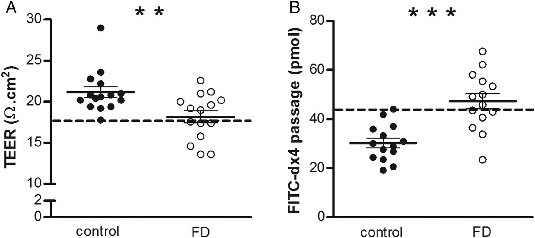
Integrity of duodenal mucosal barrier. Intestinal integrity of healthy controls (black dots) and patients with FD (white dots) was evaluated in Ussing chambers by measuring TEER (A) and passage of FITC-dx4 (B) of four duodenal biopsy specimens per subject. (A) TEER was recorded every 30 min over 2 h, and the average of all time points of the four specimens was taken. The dotted line represents the 5th centile of the control group. n=15 for both groups. (B) FITC-dx4, a probe for paracellular passage, was added to the mucosal compartment, and serosal samples were collected every 30 min over 2 h. The average of the fluorescence level at time points 60, 90 and 120 min of the four biopsy samples was taken. The dotted line represents the 95th centile of the control group. n=14 for both groups. Data are mean±SEM; **p<0.01, ***p<0.001. FD, functional dyspepsia; FITC-dx4, fluorescently labelled dextran; TEER, transepithelial electrical resistance.
Altered expression of cell-to-cell adhesion proteins at each level of the intercellular junction
As we observed that patients with FD display decreased duodenal mucosal integrity, we investigated whether altered expression of cell-to-cell adhesion proteins may underlie this phenomenon. The tight junction proteins, CLDN1–4, OCLN and ZO-1–3, the adherens junction proteins, β-catenin and E-cadherin, and the desmosomal proteins, DSC2 and DSG2, were analysed using real-time RT-PCR, western blotting and/or immunofluorescence.
Tight junction
No difference in protein expression of CLDN3–4 (p>0.10 for both) was observed in patients with FD (figure 2A,B). Expression of ZO-1 at the tight junction was significantly lower in patients than in controls (1019 (730–2066) vs 2226 (1108–3313) arbitrary fluorescence units, p=0.03) (figure 2C,D), but no redistribution to the intracellular compartment was detected. In addition, patients with FD displayed decreased gene (table 3) and protein (0.72-fold, p=0.02) expression of OCLN, with reduced phosphorylation of serine/threonine residues (p-OCLN) (0.44-fold, p=0.004) (figure 2A,B). The difference in protein expression of p-OCLN between patients with FD and healthy volunteers persisted after correction for multiple testing (p=0.02), but the difference in gene expression of OCLN and protein expression of ZO-1 and OCLN did not.
Gene expression of the cell-to-cell adhesion proteins

Expression of tight junction proteins in the duodenal mucosa. CLDN3-4, OCLN and p-OCLN expression was evaluated by western blot (A, B) and ZO-1 expression by immunofluorescence (C, D) in healthy volunteers (black dots) and patients with FD (white dots). (A) Results were normalised to the housekeeping protein, vinculin, and protein fold change was determined relative to the mean value of the control group. Data are mean±SEM; n=15 for both groups. (B) Representative western blot of six controls and six patients with FD. (C) ZO-1 expression was quantified by measuring the average fluorescence intensity in 10 non-overlapping fields per subject by confocal microscopy. Data are median (IQR); n=10 for controls and n=12 for patients with FD. (D) Representative confocal microscopy image of ZO-1 (green) in mucosal biopsy samples obtained from a control (left) and a patient (right). Scale bars: 50 µm. *p<0.05, **p<0.01. CLDN, claudin; FD, functional dyspepsia; OCLN, occludin; p-OCLN, serine/threonine-phosphorylated occludin; ZO-1, zonula occludens 1.
Adherens junction
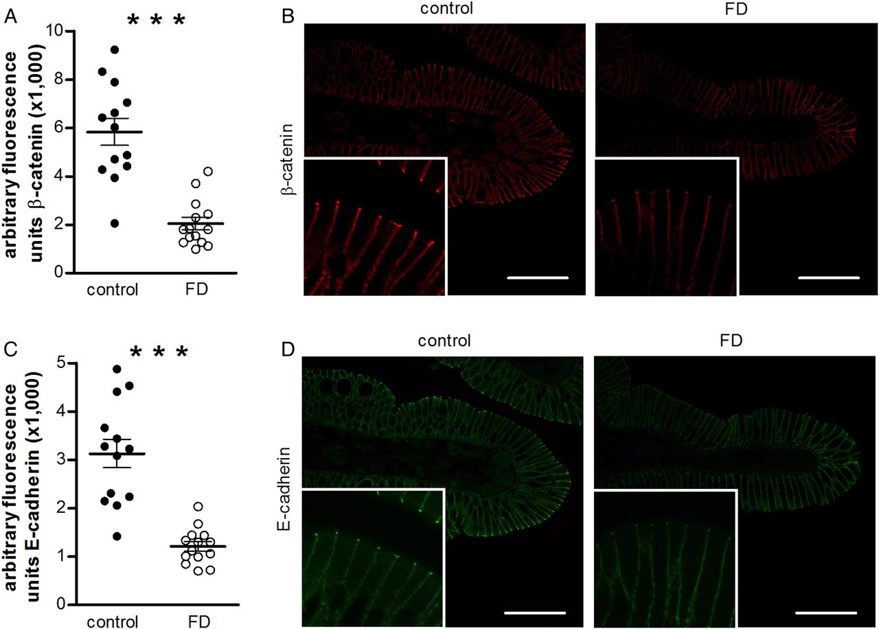
Compared with healthy volunteers, gene expression of β-catenin was decreased in patients with FD (table 3). Immunofluorescence analysis showed reduced staining of β-catenin (1800 (1275–2540) vs 6030 (4352–7472) arbitrary fluorescence units, p<0.0001) (figure 3A,B) and E-cadherin (1214±99 vs 3130±295 arbitrary fluorescence units, p<0.0001) (figure 3C,D) at the adherens junction in patients with FD. No redistribution to the cytoplasm was seen. The difference in protein expression of β-catenin (p=0.0004) and E-cadherin (p<0.0001), but not the difference in gene expression of β-catenin, between the two groups remained significant after correction for multiple testing.

Expression of adherens junction proteins in the duodenal mucosa. β-Catenin (A, B) and E-cadherin (C, D) expression was measured by immunofluorescence in healthy volunteers (black dots) and patients with FD (white dots). β-Catenin (A) and E-cadherin (C) expression was quantified by measuring the average fluorescence intensity in 10 non-overlapping fields per subject by confocal microscopy. Data are mean±SEM for E-cadherin and median (IQR) for β-catenin; n=13 for controls and n=14 for patients with FD. Representative confocal images of β-catenin (B, red) and E-cadherin (D, green) in mucosal biopsy samples obtained from a control (left) and a FD patient (right). Scale bars: 50 µm. ***p<0.001. FD, functional dyspepsia.
Desmosome
At mRNA level, the expression of DSC2 and DSG2 was reduced in patients compared with healthy subjects (table 3). Also, patients with FD displayed decreased protein expression of DSG2 (0.86-fold, p=0.02), but not of DSC2 (0.81-fold, p=0.18) (figure 4A,B). None of the significant results withstood multiple testing correction.

Expression of desmosomal proteins in the duodenal mucosa. DSC2 and DSG2 expression was measured by western blot in healthy volunteers (black dots) and patients with FD (white dots). (A) Results were normalised to the housekeeping protein vinculin and protein fold change was determined relative to the mean value of the control group. Data are mean±SEM; n=15 for both groups. (B) Representative western blot of six controls and six patients with FD. *p<0.05. DSC2, desmocollin-2; DSG2, desmoglein-2; FD, functional dyspepsia.
Patients with FD display increased eosinophil and mast cell infiltration
The presence of low-grade duodenal inflammation was evaluated by staining for eosinophils, mast cells and IELs. Compared with healthy volunteers, patients with FD showed increased eosinophil (28.3±3.0 vs 17.8±1.8 MBP+ cells/HPF, p=0.006) (figure 5A,B) and mast cell (48.5±3.1 vs 30.5±2.3 tryptase+ cells/HPF, p<0.0001) (figure 5C, D) counts. The number of mast cells did not correlate with the number of eosinophils (r=−0.29, p=0.16). No significant difference in IEL count between patients and controls (23.9±2.9 vs 18.5±1.1 CD3 cells/100 ECs, p=0.09) was detected.

Immune cell infiltration. Duodenal biopsy specimens from healthy volunteers (black dots) and patients with FD (white dots) were stained for eosinophils using eosinophilic MBP (A, B), for mast cells using tryptase (C, D) and for IELs using CD3 (E, F). Cells positive for MBP (n=14 for controls and n=12 for patients with FD) (A) and tryptase (n=15 for controls and n=12 for patients with FD) (C) were counted in at least seven non-overlapping HPFs per subject. The number of IELs was determined by counting at least 300 ECs (n=13 for controls and n=12 for patients with FD) (E). Data are mean±SEM. Representative images of MBP (B), tryptase (D) and CD3 (F) immunohistochemistry in mucosal biopsy samples obtained from a control (left) and a patient (right). Scale bar: 20 µm (B, D) or 30 µm (F). **p<0.01, ***p<0.001. EC, epithelial cell; FD, functional dyspepsia; HPF, high power field; IEL, intraepithelial lymphocyte; MBP, major basic protein.
Expression of cell-to-cell adhesion proteins correlates with duodenal integrity and low-grade inflammation
We studied the association between expression of the cell-to-cell adhesion proteins that remain significantly different between patients with FD and controls after correction for multiple testing (p-OCLN, β-catenin and E-cadherin), and measures of permeability (TEER and FITC-dx4 passage) and low-grade inflammation (mast cell and eosinophil counts) (table 4). The expression of p-OCLN correlated with TEER and the number of mast cells and eosinophils. In addition, the expression of β-catenin and E-cadherin correlated with FITC-dx4 passage and the number of mast cells and eosinophils. After correction for multiple testing, the correlation between the expression of p-OCLN and eosinophil counts (p=0.04) and the correlation between the expression of E-cadherin and mast cell counts (p=0.006) remained significant.
Correlation of cell-to-cell adhesion proteins with duodenal integrity and low-grade inflammation
Correlation of duodenal integrity with clinical factors
The association of TEER and FITC-dx4 passage with several clinical factors in the patient group—that is, the DSS and the score on the VSI and PHQ-9 questionnaire—was evaluated. A strong and significant negative correlation between TEER and the score on the VSI questionnaire was found (r=−0.62, p=0.03). However, this correlation did not remain significant after correction for multiple testing. There was no correlation between TEER and FITC-dx4 passage with the other clinical factors (r=0.38, p>0.20 for all).
Discussion
Our study is the first to demonstrate impaired duodenal mucosal barrier function in patients with FD. This is illustrated by reduced TEER, increased paracellular passage, and altered expression of several cell-to-cell adhesion proteins compared with healthy volunteers. Moreover, patients with FD are characterised by increased infiltration of mucosal mast cells and eosinophils, indicating the presence of low-grade inflammation. Furthermore, the altered expression of certain cell-to-cell adhesion proteins correlates with impaired duodenal integrity and low-grade inflammation. A summary of our findings is shown in figure 6.

Summary of results. Our data indicate the presence of decreased duodenal mucosal integrity in patients with FD. Functional evidence of increased permeability is provided, and altered expression of several cell-to-cell adhesion proteins was observed. We also showed that patients with FD were characterised by low-grade inflammation, demonstrated by increased numbers of mast cells and eosinophils. Impaired barrier function can facilitate the passage of luminal antigens through the epithelium, by which they can enter the lamina propria. This may lead to low-grade inflammation, which can then contribute to the generation and persistence of gastrointestinal symptoms. CLDN, claudin; DSC2, desmocollin-2; DSG2, desmoglein-2; FD, functional dyspepsia; IEL, intraepithelial lymphocyte; OCLN, occludin; ZO-1, zonula occludens 1.
Several reports point towards involvement of the duodenum in the pathophysiology of FD, which provided the rationale to use duodenal biopsy specimens in our study.17 Moreover, recent studies found evidence for the presence of duodenal low-grade inflammation. Our group showed the presence of focal lymphoid aggregates and increased numbers of macrophages in the duodenum of post-infectious patients with FD.5 Others reported elevated numbers of eosinophils and macrophages in the duodenum in FD.6–8 In concordance with previous reports,5 ,8 our cohort of patients with FD displayed no increase in IELs. We did detect increased infiltration of eosinophils and mast cells into the duodenal mucosa, providing support for an inflammatory mechanism in the pathogenesis of FD. The possible pathophysiological involvement of low-grade inflammation has previously been addressed in IBS, which is—together with FD—the most common functional gastrointestinal disorder. At least a subset of patients with IBS is characterised by an increased presence of immune cells such as IELs and mast cells in jejunal and colonic biopsy specimens.18 ,19 The reason for low-grade inflammation in functional gastrointestinal disorders is unclear, but impaired intestinal barrier function has been suggested to play a key role. In fact, several studies have described increased intestinal permeability in IBS subpopulations.9 Moreover, impairment of the epithelial barrier is not only present in functional gastrointestinal disorders, but is commonly found in various gastrointestinal diseases with marked intestinal inflammation, such as Crohn's disease, ulcerative colitis and coeliac disease.10 We evaluated whether increased intestinal permeability also has potential to be a pathogenic mechanism in FD. Since there is no in vivo test available that specifically measures duodenal permeability, we decided to use an ex vivo model with duodenal biopsy samples mounted in Ussing chambers. Our present study is of particular interest, as it demonstrates for the first time that mucosal integrity is impaired in patients with FD, raising the possibility that restoration of barrier function may be an attractive therapeutic target.
Intestinal barrier function is dependent on the connection of adjacent cells through junctional complexes at sites of cell–cell contact, consisting of tight junctions, adherens junctions and desmosomes. Aberrant expression of tight junction proteins seems to be implicated in intestinal permeability disorders.19–31 The tight junction barrier has at least two transport routes.32 The pore pathway is a size- and charge-selective high-capacity route for ions that is thought to be regulated by CLDNs. Since no changes in expression of the CLDNs were observed in our study, it is possible that the pore pathway is not involved in the increased permeability in patients with FD. However, we cannot exclude the possibility that CLDNs other than the ones we analysed are affected. Recent findings have shown that OCLN and ZO-1 are implicated in the regulation of the leak pathway, which is a low-capacity route that has no charge selectivity and allows limited passage of larger molecules.33 On the basis of the abnormal expression of OCLN and ZO-1 in our cohort of patients, we conclude that the leak pathway is affected in FD. Defects in this pathway may facilitate the passage of dietary antigens and bacterial products through the epithelial barrier, via which they can enter the lamina propria and possibly induce inflammatory changes.32 Besides reduced expression of OCLN, we also detected decreased serine/threonine phosphorylation of the protein. In general, phosphorylation and dephosphorylation of OCLN on serine/threonine residues is associated with assembly and disassembly, respectively, of the tight junction.34 ,35 Hence, deregulated phosphorylation may help to explain the increased duodenal permeability seen in our patient group.
We are the first to evaluate cell-to-cell adhesion proteins at each level of the intercellular junction in the same cohort. Defects in the integrity of the adherens junction have been proposed to underlie the increased mucosal permeability in Crohn's disease and ulcerative colitis.36 ,37 To the best of our knowledge, the expression of desmosomal proteins in intestinal permeability disorders has not been previously examined. An in vitro study showed that an antibody directed against DSG2 decreased TEER and increased paracellular passage, suggesting that DSG2-mediated adhesion is involved in the maintenance of the intestinal barrier.38 The reduced expression of β-catenin, E-cadherin and DSG2 that we observed in patients with FD can therefore be another mechanism contributing to impaired duodenal integrity. Further studies are, however, required to define the precise role of these proteins in intestinal barrier function. Adherens junction proteins are important not only for connecting neighbouring cells, but also for regulating cellular polarisation, apoptosis, proliferation and differentiation.39 Also, DSG2 seems to play a role in the regulation of cell viability.40 This opens the possibility that the decreased expression of these proteins in patients with FD has other effects besides increasing intestinal permeability.
Another remarkable finding of our study was that the expression of p-OCLN, β-catenin and E-cadherin correlated with duodenal integrity and low-grade inflammation. These data suggest that altered expression of cell-to-cell adhesion proteins underlies the increased permeability and that this is linked to low-grade inflammation. On the basis of this observation, we can hypothesise that a mechanistic association exists between impaired mucosal integrity and low-grade inflammation; however, this correlation should be interpreted with caution since a causal relationship remains to be established. Although we do not know whether increased permeability is the cause or the consequence of low-grade inflammation, evidence points towards a close relationship between impaired integrity and inflammatory activity. Research in animal models of intestinal inflammation has shown that restoring the intestinal barrier prevents development of inflammation.41 ,42 Moreover, eosinophil-derived corticotropin-releasing hormone can activate mast cells to decrease colonic43 and jejunal44 epithelial barrier function. Also, in patients with Crohn's disease, immune activation correlates with increased intestinal permeability, and improvement in intestinal inflammation tightens the intestinal barrier.45 ,46
We could not demonstrate a significant correlation between the symptom score of the patients with FD and duodenal integrity, which contrasts with earlier findings in patients with IBS.19 ,47 It should be mentioned that the severity of IBS can be assessed by validated questionnaires, in combination with evaluation of bowel movements and stool consistency, while similar well-established tools for quantifying the severity of FD are currently lacking. Moreover, larger studies need to be performed to disprove or confirm this result. Interestingly, we found a link between a psychological and a physiological factor in patients with FD, since we demonstrated a correlation between gastrointestinal-specific anxiety (VSI score) and TEER. But caution must be applied when interpreting this result in terms of cause and effect. This correlation did not survive correction for multiple testing, but it is of high magnitude, making it highly likely that the loss of significance is due to the low n value in this analysis.
The FD population we studied showed some heterogeneity in terms of IBS comorbidity, depression scores and the use of acid-suppressive therapy. However, the variables are in line with the clinical and epidemiological characteristics of FD,3 ,12 ,48 and our patient group all presented primarily with upper gastrointestinal symptoms that were diagnosed as FD. We have already reported that the presence of coexisting IBS had a minor effect on symptom pattern and plausible pathophysiological mechanisms in FD,48 which also seems to be the case in this study since there was no difference in permeability measurements between IBS-positive and IBS-negative patients with FD. Furthermore, only two patients presented with coexisting major depressive disorder13 and none of the patients were taking NSAIDs, corticosteroids or other immunosuppressive drugs. In addition, although the patients with FD had a lower BMI than the controls, it was within the normal range in both groups. Most importantly, the significant difference in TEER and FITC-dx4 passage between the two groups persisted after correction for all these factors, and no association between these factors and the permeability measurements was found.
In conclusion, our findings indicate for the first time that impaired duodenal barrier function may represent a pathophysiological mechanism in FD. Not only do we present functional evidence of increased permeability in patients with FD, we also provide clear structural evidence embodied by altered expression of cell-to-cell adhesion proteins. Further exploration of the processes that lead to impairment of the intestinal barrier may therefore be important for the development of therapeutic strategies. In addition, we confirmed the presence of low-grade inflammation in these patients and demonstrated that this correlated with changes in duodenal mucosal integrity. Together, these findings challenge the classical paradigm that the structure of the gastrointestinal tract is unaltered in FD.
Acknowledgments
We thank all volunteers and patients for their participation in the study. We also thank the study nurses of the Gastrointestinal Motility Unit, Leuven University Hospitals, and our colleagues of the Digestive Disease Research Unit in Barcelona, especially Cesar Sevillano, for excellent technical work. Confocal imaging was performed in the Cell Imaging Core (KU Leuven, Belgium).
References
Supplementary materials
Supplementary Data
This web only file has been produced by the BMJ Publishing Group from an electronic file supplied by the author(s) and has not been edited for content.
Files in this Data Supplement:
- Data supplement 1 - Online figure
- Data supplement 2 - Online tables
Footnotes
-
Contributors HV: study concept and design; acquisition of data; analysis and interpretation of data; drafting of the manuscript. MV: study concept and design; obtained funding; critical revision of the manuscript for important intellectual content. TV: study concept and design; critical revision of the manuscript for important intellectual content. LVO: critical revision of the manuscript for important intellectual content; statistical analysis. CM: technical support. ÅVK: technical support. NP: technical support. JS: obtained funding; material support. JDS: technical support. JT: study concept and design; patient recruitment; critical revision of the manuscript for important intellectual content; obtained funding; study supervision. RF: study concept and design; analysis and interpretation of data; obtained funding; critical revision of the manuscript for important intellectual content; study supervision
-
Funding Provided by a Methusalem grant from Leuven University to JT and by the Research Foundation Flanders (Fonds Wetenschappelijk Onderzoek Vlaanderen, FWO) through research grants (G.0863.1; to JT and RF) (KAN2012 1.5.155.12; to RF) and by doctoral (to HV and TV) and post-doctoral (to LVO and RF) fellowships. Funding was also provided by the Swedish Research Council—Medicine (to JDS), the Fondo de Investigación Sanitaria and CIBERehd, Instituto Carlos III, Subdirección General de Investigación Sanitaria, Ministerio de Ciencia e Innovación (CP10/00502 to MV; PI08/0940 and PI11/00716 and CB06/04/0021 to JS) and the 2010 Rome Foundation Award (to JS). CIBERehd is funded by the Instituto de Salud Carlos III.
-
Competing interests None.
-
Ethics approval Leuven University Hospital Ethics Committee.
-
Patient consent Obtained.
-
Provenance and peer review Not commissioned; externally peer reviewed.