Article Text

Abstract
Black esophagus, also known as acute esophageal necrosis (AEN) syndrome, is a rare entity characterized by patchy or diffuse circumferential black pigmentation of the esophageal mucosa from ischemic necrosis. It may present with life-threatening upper gastrointestinal hemorrhage resulting in high mortality in immunocompromised patients. Advanced age with multiple comorbidities compounded with compromised hemodynamic states are poor prognostic factors. Findings on laboratory work-up and radiological imaging are non-specific. After initial resuscitation, endoscopic evaluation and histological examination of esophageal biopsy are diagnostic. Early recognition and aggressive resuscitation are the fundamental principles for the management of AEN and better outcome of the disease. We report a case of a 56-year-old woman with diabetes mellitus, gastro-esophageal reflux disease, and active alcohol binging who presented with hematemesis and acute epigastric pain due to AEN. This case illustrates a rare etiology of AEN due to active alcohol drinking, which may be overlooked. Physician awareness about this etiology is important as early recognition and timely management may improve survival.
- oesophageal lesions
- gastro-oesophageal reflux disease
- endoscopy
- alcohol-induced injury
- erosive oesophagitis
This is an open access article distributed in accordance with the Creative Commons Attribution Non Commercial (CC BY-NC 4.0) license, which permits others to distribute, remix, adapt, build upon this work non-commercially, and license their derivative works on different terms, provided the original work is properly cited, appropriate credit is given, any changes made indicated, and the use is non-commercial. See: http://creativecommons.org/licenses/by-nc/4.0/.
Statistics from Altmetric.com
- oesophageal lesions
- gastro-oesophageal reflux disease
- endoscopy
- alcohol-induced injury
- erosive oesophagitis
Introduction
Acute esophageal necrosis (AEN) is a rare syndrome that is characterized by patchy or diffuse circumferential black appearance of the intrathoracic osophageal mucosa. AEN is also known as ‘esophageal stroke’ or ‘black esophagus’ due to the black pigmentation of the esophageal mucosa from ischemic necrosis seen on histopathology. The incidence of AEN is 0.01%–0.28%, with a total reported number of cases in the current literature less than 150.1 Recently, autopsy study on 310 cases has shown 10.3% incidence of AEN, indicating under-representation of the disease in the current literature especially in asymptomatic patients.2 Predisposing risk factors for AEN include male gender, advanced age (usually in the sixth decade of life), paraesophageal hernia, trauma, and presence of other comorbidities such as diabetes mellitus (DM), diabetic ketoacidosis, hypertension, cardiovascular disease, peripheral artery disease, atherosclerosis, thromboembolic disease, chronic kidney disease, chronic liver disease, malnutrition, chronic alcohol abuse, and postirradiation and solid organ or hematological malignancies.1 Overall, mortality can approach as high as 32%.3
Case description
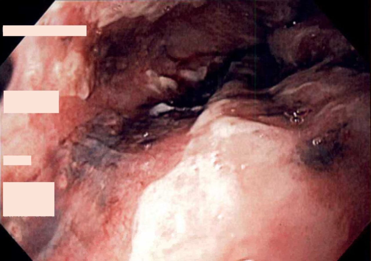
A 56-year-old woman with a history of DM, gastroesophageal reflux disease (GERD), and alcohol abuse presented to the emergency room with a 2-day history of abdominal pain and intractable nausea and vomiting for 24 hours. The abdominal pain was sharp epigastric, 10/10 in severity, and non-radiating, precipitated with ingestion of oral intake, without alleviating factors. Prior to presentation, she had more than 30 episodes of vomiting, which was initially clear, but progressed to coffee ground emesis. She had no melena or hematochezia. Her social history was consistent with alcohol abuse (8–10 beers per day), without history of smoking, or substance or domestic abuse. On examination, she was tachycardic (120 per minute) and hypotensive (80/50 mm Hg), had oropharyngeal erythema with white spots on its posterior wall, and significant ptyalism. She had epigastric tenderness and unremarkable digital rectal examination. Laboratory work-up revealed lactic acidosis (>15 mmol/L), acute kidney injury with creatinine 2.21 mg/dL, leukocytosis (18 ×109/L), transaminitis with aspartate aminotransferase 175 U/L, alanine aminotransferase 121 U/L, alkaline phosphate 84 U/L, and a total bilirubin of 1.2 mg/dL. A CT of the abdomen and pelvis without contrast was unremarkable except for paraesophageal hernia. The hospital course was complicated by hematemesis, dysphagia and melena. She was resuscitated with intravenous fluids and initially started on proton pump inhibitor (PPI), antiemetics, antibiotics and analgesics. After initial resuscitation, esophagogastroduodenoscopy (EGD) was performed which showed inflammation of the epiglottis, arytenoid cartilages, and dark mucosal pigmentation of the distal two-thirds of the esophagus with associated hiatus hernia (figure 1 and online supplementary file 1). A biopsy of the esophageal mucosa demonstrated fragments of fibrinopurulent exudate, and necrotic tissue with predominant neutrophil infiltration indicating severe inflammation (figure 2). A periodic acid Schiff stain was negative for fungal organisms. There was no evidence of Helicobacter pylori, cytomegalovirus (CMV), herpes simplex virus (HSV), Candida albicans infections and malignancy on further testing of biopsy specimen. She was managed with strict restrictions of oral intake, total parenteral nutrition, PPI, pain control with analgesic, and serial EGDs at 2 weeks (figure 3) and 3 weeks (figure 4) to assess for inflammation and improvement in necrosis. Complete resolution of esophageal necrosis was noted at 8-week EGD follow-up (figure 5). She was transitioned to oral diet, which she tolerated well.
Supplemental material
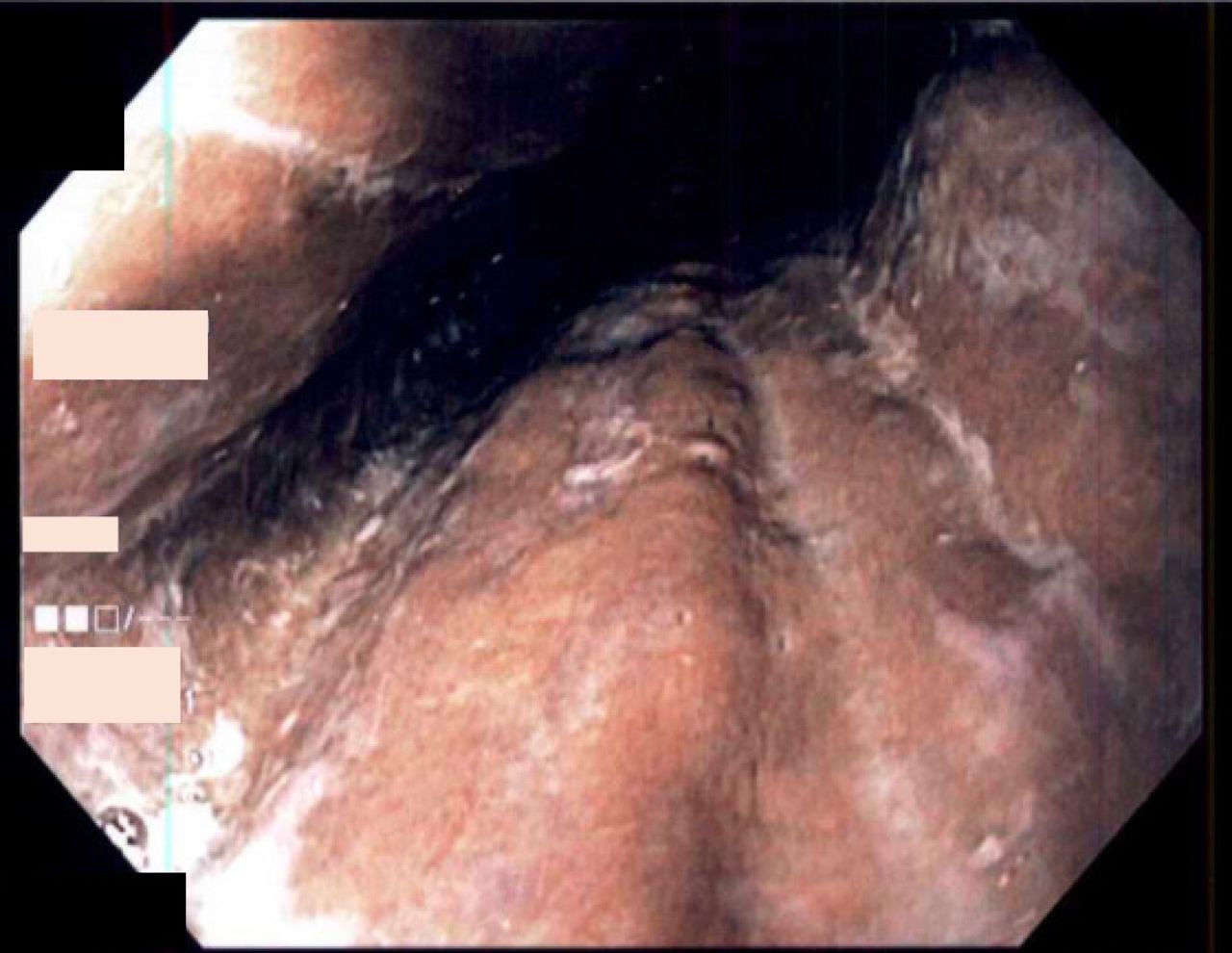
Initial upper endoscopy showing black pigmentation of esophageal mucosa due to ischemic necrosis involving distal two-thirds of the esophagus.
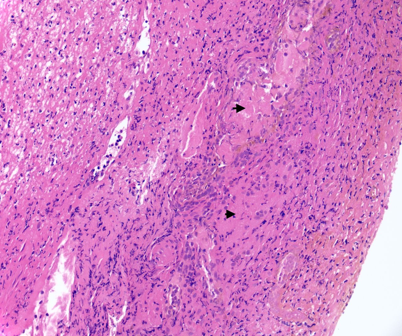
A biopsy of the esophageal mucosa. Histological examination (200X magnification) demonstrating fragments of fibrinopurulent exudate, necrotic tissue with loss of cell architectures (black arrowhead). Significant neutrophil infiltration of mucosal tissue can be seen indicating severe inflammation.

Upper endoscopy showing resolving esophageal lesions at 2 weeks.

Upper endoscopy showing resolving esophageal lesions at 3 weeks.

Follow up upper endoscopy at 8 weeks showing complete resolution of lesions.
Discussion
AEN was first reported in 1990 and was then classified as a separate syndrome in 2007.1 4 The distal third of the esophagus is commonly affected (97% of cases) due to poor vascularity; however, the length of esophageal involvement is variable depending on severity of disease and time to presentation since onset of symptoms.5
Pathogenesis and etiology
Adequate blood supply to the esophageal mucosa, lamina propria and submucosa is essential to maintain intrinsic cellular function and defence barriers. Impaired esophageal blood supply coupled with reperfusion mucosal injuries due to reactive oxygen metabolites results in direct and indirect injuries to cellular defence barriers, respectively, which cause cellular lysis and necrosis.5 As such the proposed pathophysiology of ischemic necrosis results from impairment of the mucosal barriers of the esophagus due to traumatic insult (persistent vomiting), chemical injury (from acidic contents of the stomach in the setting of GERD or gastric outlet obstruction), ongoing esophageal infections such as Klebsiella pneumoniae, CMV, and esophageal candidiasis, and hypoperfusion of the esophageal mucosa due to hemodynamic compromise.1 6 In a large systematic review of 130 patients, the commonly reported comorbidities associated with AEN are DM (38%), uncontrolled hypertension (37%), chronic alcohol abuse (25%), chronic kidney disease (16%), cardiovascular disease (10%), malignancy (10%), diabetic ketoacidosis (9%), GERD (9%), and intoxication (9%), followed by cirrhosis (9%), alcoholic hepatitis (5%), smoking (5%) and peptic ulcer disease (4%).7 In our case, the triggering etiology of AEN was binge alcohol drinking superimposed by prolonged history of DM and GERD. AEN associated with active alcohol drinking is a rare entity, and only nine cases have been reported in the current literature (table 1).8–16 Upper gastrointestinal hemorrhage, vomiting, abdominal pain and unresponsiveness are common presenting symptoms in these cases with AEN associated with drinking alcohol ranging 2–8 pints daily.8–16 These case studies suggested low systemic perfusion due to alcoholic lactic acidosis, hypothermia, and impaired mucosal defence are major pathological causes of alcohol-induced AEN.8–16 Multiple forensic studies on alcohol-induced AEN have shown hemorrhagic shock resulting from esophageal necrosis is the most common cause of death, followed by cardiac arrest and hypothermia (table 2).17–21 On autopsy, a black pigmentation involving the distal esophagus with or without Wischnewski lesions (red brownish erosions of the mucosa) and hemorrhagic fluid collection are commonly reported findings related to alcohol-induced AEN (table 2).17–21
Case studies with acute esophageal necrosis due to active alcohol drinking
Autopsy studies of alcohol-induced acute esophageal necrosis
Clinical presentation and diagnosis
The clinical presentation of AEN ranges from an asymptomatic state to a wide spectrum of clinical manifestations. Upper gastrointestinal hemorrhage is the most common clinical presentation requiring emergent hospitalization. Majority of patients present with hematemesis (66%), hypotension/syncope (36%), melena (33%), and acute abdominal pain (28%), followed by fever/infections (13%), dysphagia (12%), anemia and altered mental status or death on admission (5%).7 In severe cases, patients may present with multiorgan failure due to sepsis, acute pancreatitis, gut ischemia, acute renal failure, liver failure, coagulopathy and thromboembolism.5 22 Commonly noted findings on physical examination are cachexia, fever, hypotension, tachycardia, pallor, and abdominal tenderness; however, these findings may be confounded by the presence of multiple comorbidities.5 23 Laboratory findings such as anemia, metabolic derangement, lactic acidosis and leukocytosis are non-specific, and radiological imaging may demonstrate wall thickening of the distal esophagus, distended stomach, hiatal hernia (if present) and pneumomediastinum in case of esophageal perforation due to full thickness necrosis of the esophageal wall. EGD evaluation and histological examination of biopsy specimen are essential for diagnostic confirmation of AEN by differentiating it from esophageal pigmentation due to mucosal melanosis, pseudomembrane esophagitis, acanthosis nigricans, ingestion of coal dust or dye, and ruling out other etiologies such as malignancy, H. pylori, CMV, HSV or fungal infection.24 EGD visualizations of patchy or circumferential pigmentation of the distal esophageal mucosa with clear demarcation of the normal mucosa at the gastroesophageal junction are characteristic features of AEN.
Our patient had multiple risk factors, including DM, GERD, and prolonged vomiting after binge alcohol drinking (8–10 beers daily), which resulted in traumatic injury to the distal esophageal mucosa. She may also have impaired vascularity of the mucosa in the setting of prolonged DM and disruption of the mucosal barrier due to chemical injury from gastric contents in the presence of hiatal hernia and GERD. Although the patient presented with acute epigastric abdominal pain without hematemesis, she developed coffee ground emesis during hospitalization likely due to traumatic injury to the esophageal mucosa in the setting of retching and persistent vomiting. She further developed hemodynamic compromise in the setting of septic shock as indicated by hypotension, leukocytosis and lactic acidosis. EGD findings and histological examination of biopsy were diagnostic of AEN. Black esophagus due to mucosal necrosis progresses from distal to the proximal esophagus in the majority of cases; therefore, a meticulous EGD evaluation and biopsy of non-black (proximal end of involved) esophagus may show acute necrotizing changes without pigmentation, as the advancement of endoscopy to take biopsy of black (distal end) esophagus may put patients at high risk of esophageal perforation through friable mucosa. This is why biopsy sampling was performed at the proximal end of the black esophagus and no pigmentation was seen on histological examination of our patient.
Management
The mainstay of medical management of AEN is directed at hemodynamic stabilization with initial resuscitation, supportive care and treatment of underlying etiologies. In patients with acute upper gastrointestinal hemorrhage, aggressive management with nothing per oral, intravenous fluids, PPI, and transfusion of blood products is needed. Supportive care with antiemetics, analgesic and parenteral nutrition may be required in those with multiorgan failure and pre-existing malnutrition. Broad spectrum empirical antimicrobial therapy should be promptly started in patients with sepsis, acute decompensation, immunocompromised state, and in those who presented with complications such as esophageal perforation and multiorgan failure. An estimated 60% of patients recover with conservative management. A large case series showed development of esophageal strictures in 25% of patients with AEN due to failed medical management requiring sequential endoscopic dilation of stricture.24 The presence of coexisting duodenal ulcer is a potential risk factor associated with the development of esophageal stricture in 35%–50% of cases.24 Esophageal perforation was reported in 5% of patients, which may result in mediastinal abscess or mediastinitis in a small proportion of cases. These patients are surgically managed with mediastinal lavage, decortication of abscess with delayed repair of esophagus and occasionally with distal esophagectomy.5 Our patient was managed with initial hemodynamic resuscitation, antimicrobial therapy and supportive care with complete resolution of symptoms in 8 weeks.
Conclusion
Active alcohol drinking is a rare etiology of AEN, which may present with acute upper gastrointestinal hemorrhage. The prognosis of AEN is poor and a high index of clinical suspicion and awareness of this rare etiology is required to pursue EGD for early diagnosis and timely management. In case of circumferential necrosis of the esophagus, a meticulous EGD evaluation is required as risk of perforation is high due to transmural involvement. Early recognition and aggressive resuscitation are the fundamental principles for the management of AEN and better outcome of the disease.
References
Footnotes
Twitter @M_Nadeem_Yousaf
Contributors AS, FSC, HAN: manuscript writing, figures, tables and data review. NS, RF: proof-reading and data review. MNY: manuscript writing, revisions and overall supervision.
Funding The authors have not declared a specific grant for this research from any funding agency in the public, commercial or not-for-profit sectors.
Competing interests None declared.
Patient consent for publication Obtained.
Provenance and peer review Not commissioned; externally peer reviewed.
Data availability statement Data are available in a public, open access repository, except for patient-related data.