Article Text

Abstract
Background/aims Although percutaneous endoscopic gastrostomy (PEG) is the method of choice for long-term enteral nutrition, feeding-related adverse events such as aspiration pneumonia and peristomal leakage can impede the use of PEG. Percutaneous endoscopic transgastric jejunostomy (PEG-J) using large-bore jejunal tubes may help by circumventing gastric passage during enteral nutrition and improving drainage of gastric secretions.
Methods 20 patients (12 males and 8 females) who received PEG-J after unsuccessful PEG feeding during a 6-year period in our institution were analysed retrospectively to evaluate the efficacy of large-bore jejunal feeding tubes in maintaining enteral nutrition.
Results The median age was 83.5 (71–96) years. The median period between PEG and PEG-J was 33 (14–280) days. Indications were aspiration due to gastro-oesophageal reflux in 18 patients and severe peristomal leakage in 2 patients. Tube placements were successful in all patients. There were 6 (30%) in-hospital mortalities, with 3 (15%) occurring within 30 days after procedure.
Conclusions PEG-J can be performed safely in patients with PEG and may facilitate the maintenance of enteral nutrition in most of the patients. Patients with unsuccessful PEG feeding can be offered the option of jejunal feeding before considering termination of enteral nutrition.
- ENDOSCOPIC GASTROSTOMY
- ENTERAL NUTRITION
- NUTRITIONAL SUPPORT
This is an Open Access article distributed in accordance with the Creative Commons Attribution Non Commercial (CC BY-NC 4.0) license, which permits others to distribute, remix, adapt, build upon this work non-commercially, and license their derivative works on different terms, provided the original work is properly cited and the use is non-commercial. See: http://creativecommons.org/licenses/by-nc/4.0/
Statistics from Altmetric.com
Summary box
What is already known about this subject?
▸ Feeding-related adverse events such as aspiration pneumonia and peristomal leakage can impede gastric feeding.
▸ Percutaneous endoscopic transgastric jejunostomy (PEG-J) circumvents gastric passage during enteral nutrition and can provide drainage for excessive gastric secretions.
What are the new findings?
▸ After unsuccessful percutaneous endoscopic gastrostomy, jejunal feeding via PEG-J helps maintain enteral nutrition in most patients.
▸ However, feeding intolerance still persists in some patients as not all patients benefit from the procedure.
How might it impact on clinical practice in the foreseeable future?
▸ Patients with unsuccessful PEG feeding can be offered the option of jejunal feeding before considering termination of enteral nutrition.
Introduction
The introduction of percutaneous endoscopic gastrostomy (PEG) provided a safe and minimally invasive procedure for long-term enteral nutrition in patients with dysphagia or insufficient oral intake.1–3 However, feeding-related adverse events such as aspiration pneumonia due to gastro-oesophageal reflux of gastric feed and uncontrolled peristomal leakage can impede the use of PEG. Although jejunal (or postpyloric) feeding has not been established as being superior to gastric feeding,4–6 it may help overcome gastric feeding-related adverse events by circumventing gastric passage during enteral nutrition.7–9 Jejunal feeding can be achieved by direct percutaneous endoscopic jejunostomy (D-PEJ)10 or more commonly by placing a jejunal tube through an existing gastrostomy site, also referred to as percutaneous endoscopic transgastric jejunostomy (PEG-J or jejunal tube through PEG).11 Jejunal extension tubes placed through PEG tubes are usually smaller and longer compared to tubes used in D-PEJ, making them more prone to tube dysfunctions such as obstruction or migration into the stomach.12 ,13 Instead of jejunal extension tubes, large-bore jejunal tubes can also be placed directly via the PEG puncture site with the aid of an ultrathin endoscope after removal of the PEG tube.14 This study analyses our experience with PEG-J using large-bore jejunal tubes with gastric decompression function (providing simultaneous drainage of gastric secretions) in patients with unsuccessful PEG feeding.
Methods
Study design and patients
Medical records of patients who received PEG tube placements in our hospital between January 2007 and December 2012 were screened. Patients who were not discharged in a timely manner due to feeding-related adverse events and subsequently underwent PEG-J to maintain enteral nutrition were enrolled in our study and reviewed retrospectively. Patients who were discharged once or had their PEG/PEG-J tube placed for bowel decompression were excluded from the study. Data regarding baseline characteristics such as age, gender, comorbidities and preoperative biomarkers (body mass index and blood laboratory markers) as well as postprocedural clinical outcomes (length of stay and mortality) were studied. A 20 Fr size all-silicone jejunal tube (effective length 40 cm) with gastric decompression function (figure 1; Cliny PEG-J Catheter by Create Medic Co., Ltd, Yokohama, Japan) was used and all procedures were performed in an interventional radiology suite with the use of fluoroscopy. This study was reviewed and approved by the Institutional Ethics Review Board of Hiroshima Kyoritsu Hospital. All patients, or their legal guardians, provided informed written consent before undergoing procedures.

Percutaneous endoscopic transgastric jejunostomy tube used in our hospital.
PEG-J tube insertion technique
Existing PEG tubes were removed and an ultrathin endoscope (5.4 mm outer diameter) was inserted into the gastric lumen through the gastrostomy puncture site (figure 2A, red arrow). Once the endoscope was fluoroscopically confirmed to have passed the ligament of Treitz, a guide wire was inserted and the endoscope removed (figure 2B). With the aid of the guide wire, the PEG-J tube was then placed so that the tip of the tube is located in the jejunum (figure 2C). After removal of the guide wire, procedural success was confirmed using a contrast medium (figure 2D). Tube feeding usually resumed the following day by gravity-controlled drip feeding adjusted to about 100 mL/hour with or without a pump.
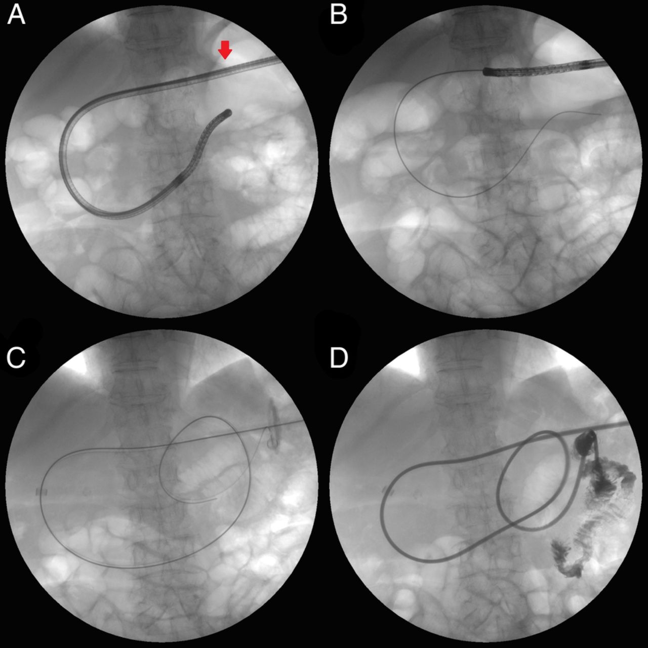
Radiological imaging of percutaneous endoscopic transgastric jejunostomy tube placement.
Statistical analysis
Continuous variables are expressed either as a median (range) or as a mean (SD). Categorical variables are expressed as numbers (percentage). Comparisons for continuous variables, considered non-parametric, in the same group were made using the Wilcoxon matched-pairs signed ranks test. Statistical significance was defined as p<0.05 and analysis was performed using XLSTAT2014 for Windows (Addinsoft Ltd., Paris, France).
Results
Twenty patients (age 71–96 years, 12 men) met the criteria for enrolment. Indications for PEG-J were aspiration from gastric feed reflux (confirmed by clinical symptoms and radiological findings) in 18 patients and severe peristomal leakage in 2 patients. Tube placement was successful in all patients. Table 1 shows the clinical characteristics of patients who underwent the procedure. Semisolid feeds, which may improve or prevent PEG-related adverse events,15 ,16 were attempted in 13 patients (65%) before PEG-J. The period between PEG tube placements and PEG-J ranged from 14 to 280 days (median 33 days).
Clinical characteristics of patients
Patients undergoing the procedure were generally in poor condition with a mean body mass index of 17.5 kg/m2 and serum albumin levels below 3.0 g/dL (significantly lower compared to pre-PEG levels).
The postprocedural clinical course is summarised in table 2. The postprocedural hospital length of stay ranged from 7 to 289 days (median 29.5 days). Fourteen patients were discharged alive and six patients died due to aspiration pneumonia and other comorbidities. Three patients died within 30 days after PEG-J. Two patients had enteral nutrition terminated and transited to total parenteral nutrition (TPN). However, these two were also in the in-hospital mortality group. In surviving patients, the frequency of feeding-related adverse events improved and regular tube replacement was performed every 4–6 months to avoid tube dysfunction.
Postprocedural clinical course after PEG-J tube placement
Discussion
For patients with impaired oral intake, tube feeding is the alternative choice for enteral nutrition. Tube feeding can be initiated using nasogastric tubes (or nasojejunal tubes), but for the long-term, percutaneous routes are preferable.1–3 Although the most common percutaneous route is through PEG, feeding-related adverse events such as aspiration from gastro-oesophageal reflux of feed and peristomal leakage can impede enteral feeding. Administering semisolid feeds or blended food instead of conventional liquid feed may help reduce the incidence of gastro-oesophageal reflux.15 ,16 Recently, using elemental diet was shown to be effective in preventing gastric feeding-related adverse events.17
Since jejunal (or postpyloric) feeding has not been established as being superior to gastric feeding, it is not usually considered the first choice when initiating percutaneous tube feeding.4–6 The procedure (D-PEJ or PEG-J) is more complicated and long-term jejunal feeding has been associated with deficiency in micronutrients such as copper.18 Jejunal feeding via D-PEJ has also been associated with high peristomal leakage rates.19 However, by circumventing the gastric passage during enteral nutrition and improving the drainage of gastric secretions via decompression holes, jejunal feeding through PEG-J could help improve feeding-related adverse events encountered during PEG feeding.
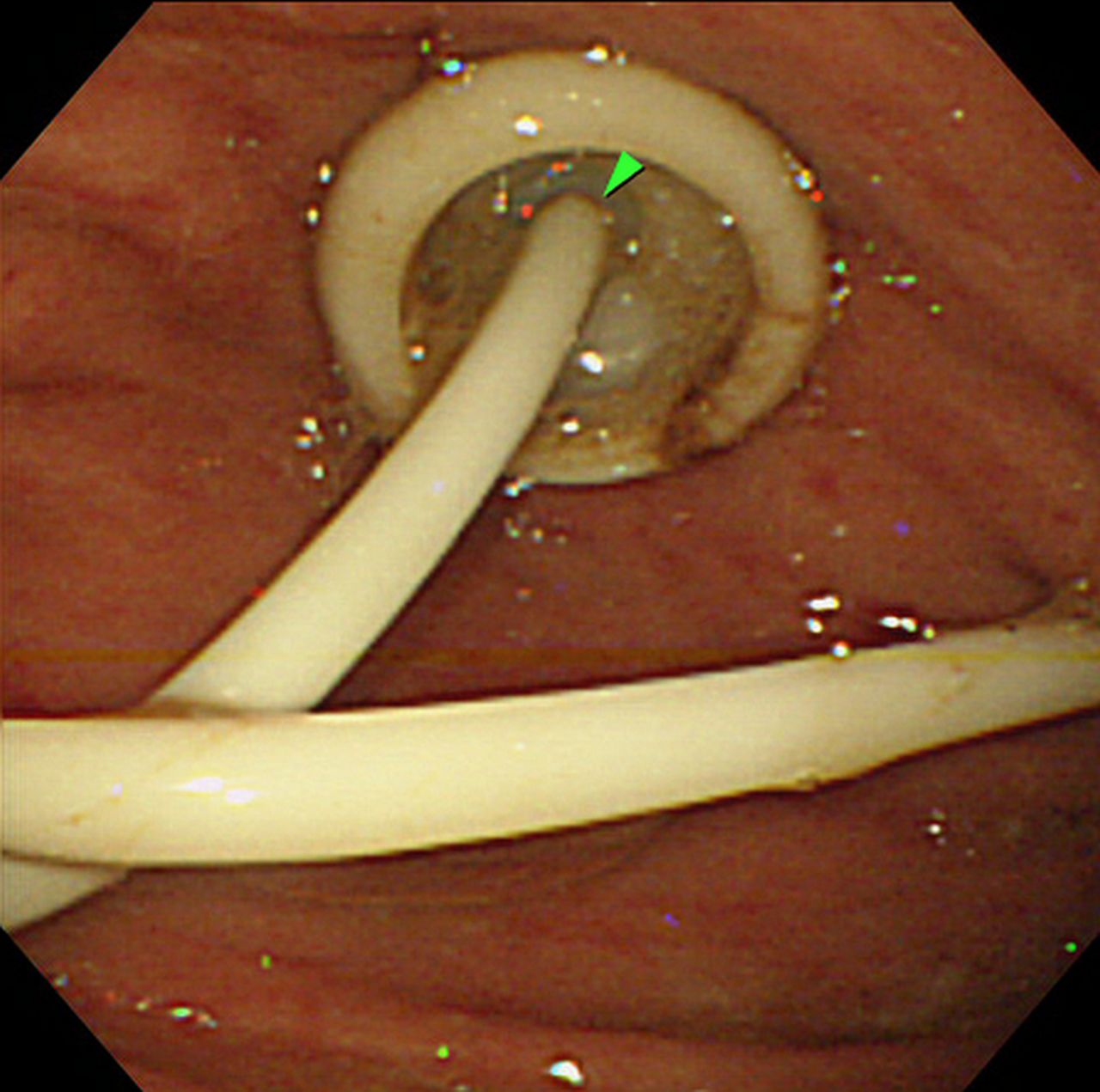
Although percutaneous jejunal feeding can be achieved by either D-PEJ or PEG-J, previous studies showed that PEG-J using jejunal extension tubes placed through PEG tubes were prone to tube dysfunction such as tube blockage or migration because of their smaller size (up to 9 Fr).12 ,13 Gastric decompression was also limited due to the almost total occlusion of the existing PEG tube's lumen (figure 3, green arrowhead). Nevertheless, for patients with PEG, accessing the jejunum through a new puncture site via D-PEJ is also not an attractive option, not to mention that D-PEJ may not be technically feasible in up to 38% of patients.20
Endoscopic image of percutaneous endoscopic transgastric jejunostomy with a jejunal extension tube through the percutaneous endoscopic gastrostomy tube.
Currently, PEG-J can also be performed using large-bore gastrojejunal tubes (up to 24 Fr size) which are placed directly through the PEG site with or without the aid of endoscopy.14 ,21 The larger tubes should theoretically reduce the frequency of tube dysfunction and tubes with gastric decompression holes, like the ones used in our hospital, also provide an outlet for excessive gastric secretions during jejunal feeding. To the best of our knowledge, there are no studies comparing D-PEJ and PEG-J using jejunal tubes more than 20 Fr size. Tube dysfunction can also be avoided or greatly reduced by regular (every 4–6 months) tube replacement as practised in Japan.
In this study, we retrospectively reviewed our experience using large-bore 20 Fr size jejunal tubes with gastric decompression function. Successful tube placement in all attempts showed agreement with previous studies citing higher technical success with PEG-J compared to D-PEJ.12 ,13 Despite the declining prognostic status as indicated by lower serum albumin levels compared to pre-PEG levels, 14 of 20 patients experienced improvement in enteral feeding and were successfully discharged without further intervention. Although transition from enteral to parenteral nutrition is an option when faced with tube feeding-related adverse events, this may also increase the cost of nutritional therapy.22 Patients in our study who transited to TPN also did not fare well due to their poor status. Limitations of this study include the small enrolment size and retrospective design.
Conclusion
PEG-J using 20 Fr size jejunal tubes with gastric decompression function can be performed safely in patients with PEG. Although it does not resolve tube feeding-related adverse events in all patients, it may help maintain enteral feeding in many patients who would otherwise be indicated for TPN. Since enteral nutrition is the route of choice as long as gut integrity is intact, PEG-J is an alternative worth exploring before terminating enteral nutrition when PEG feeding is unsuccessful.
Acknowledgments
The authors thank CREATE MEDIC Co., Ltd for providing the unannotated picture for figure 1.
References
Footnotes
Contributors EWTY designed and performed the study, analysed the data, and drafted the manuscript. EWTY, SN and KN participated in the procedures described in the study. KY assisted in the clinical data management. All authors read and approved the final manuscript.
Competing interests None declared.
Patient consent Obtained.
Ethics approval Hiroshima Kyoritsu Hospital Ethics Review Committee.
Provenance and peer review Not commissioned; externally peer reviewed.
Data sharing statement No additional data are available.